7
views
0
recommends
+1 Recommend
0 collections
    0
    shares
      • Record: found
      • Abstract: found
      • Article: found
      Is Open Access

      Brain network disintegration as a final common pathway for delirium: a systematic review and qualitative meta-analysis

      review-article

      Read this article at

      Bookmark
          There is no author summary for this article yet. Authors can add summaries to their articles on ScienceOpen to make them more accessible to a non-specialist audience.

          Abstract

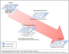
          Delirium is an acute neuropsychiatric syndrome characterized by altered levels of attention and awareness with cognitive deficits. It is most prevalent in elderly hospitalized patients and related to poor outcomes. Predisposing risk factors, such as older age, determine the baseline vulnerability for delirium, while precipitating factors, such as use of sedatives, trigger the syndrome. Risk factors are heterogeneous and the underlying biological mechanisms leading to vulnerability for delirium are poorly understood. We tested the hypothesis that delirium and its risk factors are associated with consistent brain network changes. We performed a systematic review and qualitative meta-analysis and included 126 brain network publications on delirium and its risk factors. Findings were evaluated after an assessment of methodological quality, providing N=99 studies of good or excellent quality on predisposing risk factors, N=10 on precipitation risk factors and N=7 on delirium. Delirium was consistently associated with functional network disruptions, including lower EEG connectivity strength and decreased fMRI network integration. Risk factors for delirium were associated with lower structural connectivity strength and less efficient structural network organization. Decreased connectivity strength and efficiency appear to characterize structural brain networks of patients at risk for delirium, possibly impairing the functional network, while functional network disintegration seems to be a final common pathway for the syndrome.

          Highlights

          • Delirium is consistently associated with functional network impairments.

          • Risk factors are associated with lower structural connectivity strength.

          • Risk factors are associated with a less efficient structural network organization.

          • Structural impairments make the functional network more vulnerable to deterioration.

          • Functional network disintegration seems to be a final common pathway for delirium.

          Related collections

          Most cited references129

          • Record: found
          • Abstract: found
          • Article: not found
          Is Open Access

          Benchmarking of participant-level confound regression strategies for the control of motion artifact in studies of functional connectivity.

          Since initial reports regarding the impact of motion artifact on measures of functional connectivity, there has been a proliferation of participant-level confound regression methods to limit its impact. However, many of the most commonly used techniques have not been systematically evaluated using a broad range of outcome measures. Here, we provide a systematic evaluation of 14 participant-level confound regression methods in 393 youths. Specifically, we compare methods according to four benchmarks, including the residual relationship between motion and connectivity, distance-dependent effects of motion on connectivity, network identifiability, and additional degrees of freedom lost in confound regression. Our results delineate two clear trade-offs among methods. First, methods that include global signal regression minimize the relationship between connectivity and motion, but result in distance-dependent artifact. In contrast, censoring methods mitigate both motion artifact and distance-dependence, but use additional degrees of freedom. Importantly, less effective de-noising methods are also unable to identify modular network structure in the connectome. Taken together, these results emphasize the heterogeneous efficacy of existing methods, and suggest that different confound regression strategies may be appropriate in the context of specific scientific goals.
            Bookmark
            • Record: found
            • Abstract: found
            • Article: not found

            Structural insights into aberrant topological patterns of large-scale cortical networks in Alzheimer's disease.

            Recent research on Alzheimer's disease (AD) has shown that cognitive and memory decline in this disease is accompanied by disrupted changes in the coordination of large-scale brain functional networks. However, alterations in coordinated patterns of structural brain networks in AD are still poorly understood. Here, we used cortical thickness measurement from magnetic resonance imaging to investigate large-scale structural brain networks in 92 AD patients and 97 normal controls. Brain networks were constructed by thresholding cortical thickness correlation matrices of 54 regions and analyzed using graph theoretical approaches. Compared with controls, AD patients showed decreased cortical thickness intercorrelations between the bilateral parietal regions and increased intercorrelations in several selective regions involving the lateral temporal and parietal cortex as well as the cingulate and medial frontal cortex regions. Specially, AD patients showed abnormal small-world architecture in the structural cortical networks (increased clustering and shortest paths linking individual regions), implying a less optimal topological organization in AD. Moreover, AD patients were associated with reduced nodal centrality predominantly in the temporal and parietal heteromodal association cortex regions and increased nodal centrality in the occipital cortex regions. Finally, the brain networks of AD were about equally as robust to random failures as those of controls, but more vulnerable against targeted attacks, presumably because of the effects of pathological topological organization. Our findings suggest that the coordinated patterns of cortical morphology are widely altered in AD patients, thus providing structural evidence for disrupted integrity in large-scale brain networks that underlie cognition. This work has implications for our understanding of how functional deficits in patients are associated with their underlying structural (morphological) basis.
              Bookmark
              • Record: found
              • Abstract: found
              • Article: not found

              Disrupted brain connectivity networks in drug-naive, first-episode major depressive disorder.

              Neuroimaging studies have shown that major depressive disorder (MDD) is accompanied by structural and functional abnormalities in specific brain regions and connections; yet, little is known about alterations of the topological organization of whole-brain networks in MDD patients. Thirty drug-naive, first-episode MDD patients and 63 healthy control subjects underwent a resting-state functional magnetic resonance imaging scan. The whole-brain functional networks were constructed by thresholding partial correlation matrices of 90 brain regions, and their topological properties (e.g., small-world, efficiency, and nodal centrality) were analyzed using graph theory-based approaches. Nonparametric permutation tests were further used for group comparisons of topological metrics. Both the MDD and control groups showed small-world architecture in brain functional networks, suggesting a balance between functional segregation and integration. However, compared with control subjects, the MDD patients showed altered quantitative values in the global properties, characterized by lower path length and higher global efficiency, implying a shift toward randomization in their brain networks. The MDD patients exhibited increased nodal centralities, predominately in the caudate nucleus and default-mode regions, including the hippocampus, inferior parietal, medial frontal, and parietal regions, and reduced nodal centralities in the occipital, frontal (orbital part), and temporal regions. The altered nodal centralities in the left hippocampus and the left caudate nucleus were correlated with disease duration and severity. These results suggest that depressive disorder is associated with disruptions in the topological organization of functional brain networks and that this disruption may contribute to disturbances in mood and cognition in MDD patients. Copyright © 2011 Society of Biological Psychiatry. Published by Elsevier Inc. All rights reserved.
                Bookmark

                Author and article information

                Contributors
                Journal
                Neuroimage Clin
                Neuroimage Clin
                NeuroImage : Clinical
                Elsevier
                2213-1582
                03 April 2019
                2019
                03 April 2019
                : 23
                : 101809
                Affiliations
                [a ]Department of Intensive Care Medicine and Brain Center Rudolf Magnus, University Medical Center Utrecht, Utrecht University, Heidelberglaan 100, 3584 CX Utrecht, The Netherlands
                [b ]Department of Psychiatry and Brain Center Rudolf Magnus, University Medical Center Utrecht, Utrecht University, Heidelberglaan 100, 3584 CX Utrecht, The Netherlands
                [c ]Melbourne Neuropsychiatry Center, Department of Psychiatry, Level 3, Alan Gilbert Building, 161 Barry Street, Carlton South, 3053 Victoria, University of Melbourne and Melbourne Health, Australia
                [d ]Department of Clinical Neurophysiology and MEG Center, Neuroscience Campus Amsterdam, VU University Medical Center, De Boelelaan 1117, 1081 HV Amsterdam, The Netherlands
                [e ]Faculty of Psychology, Utrecht University, Heidelberglaan 1, 3584 CS Utrecht, The Netherlands
                [f ]Faculty of Science and Technology, University of Twente, Drienerlolaan 5, 7522 NB Enschede, The Netherlands
                Author notes
                [* ]Corresponding author at: Department of Intensive Care Medicine, University Medical Center Utrecht, HP F.06.149, Postbus 85500, 3508 GA Utrecht, The Netherlands. S.J.T.vanMontfort-2@ 123456umcutrecht.nl
                Article
                S2213-1582(19)30159-7 101809
                10.1016/j.nicl.2019.101809
                6461601
                30981940
                37e6f14a-6b35-4659-a69b-901d401de58b
                © 2019 The Author(s)

                This is an open access article under the CC BY-NC-ND license (http://creativecommons.org/licenses/by-nc-nd/4.0/).

                History
                : 1 February 2019
                : 25 March 2019
                : 31 March 2019
                Categories
                Review Article

                delirium,brain,networks,connectome,aging,cognitive impairment
                delirium, brain, networks, connectome, aging, cognitive impairment

                Comments

                Comment on this article