74
views
0
recommends
+1 Recommend
0 collections
    0
    shares
      • Record: found
      • Abstract: found
      • Article: found
      Is Open Access

      Clinical Manifestations of Portal Hypertension

      review-article

      Read this article at

      Bookmark
          There is no author summary for this article yet. Authors can add summaries to their articles on ScienceOpen to make them more accessible to a non-specialist audience.

          Abstract

          The portal hypertension is responsible for many of the manifestations of liver cirrhosis. Some of these complications are the direct consequences of portal hypertension, such as gastrointestinal bleeding from ruptured gastroesophageal varices and from portal hypertensive gastropathy and colopathy, ascites and hepatorenal syndrome, and hypersplenism. In other complications, portal hypertension plays a key role, although it is not the only pathophysiological factor in their development. These include spontaneous bacterial peritonitis, hepatic encephalopathy, cirrhotic cardiomyopathy, hepatopulmonary syndrome, and portopulmonary hypertension.

          Related collections

          Most cited references102

          • Record: found
          • Abstract: found
          • Article: not found

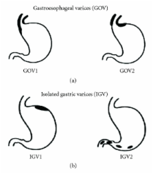
          Prevalence, classification and natural history of gastric varices: a long-term follow-up study in 568 portal hypertension patients.

          To determine the prevalence and natural history of gastric varices, we prospectively studied 568 patients (393 bleeders and 175 nonbleeders) with portal hypertension (cirrhosis in 301 patients, noncirrhotic portal fibrosis in 115 patients, extrahepatic portal vein obstruction in 117 patients and hepatic venous outflow obstruction in 35 patients). Primary (present at initial examination) gastric varices were seen in 114 (20%) patients; more were present in bleeders than in non-bleeders (27% vs. 4%, respectively; p < 0.001). Secondary (occurring after obliteration of esophageal varices) gastric varices developed in 33 (9%) patients during follow-up of 24.6 +/- 5.3 mo. Gastric varices (compared with esophageal varices) bled in significantly fewer patients (25% vs. 64%, respectively). Gastric varices had a lower bleeding risk factor than did esophageal varices (2.0 +/- 0.5 vs. 4.3 +/- 0.4, respectively) but bled more severely (4.8 +/- 0.6 vs. 2.9 +/- 0.3 transfusion units per patient, respectively). Once a varix bled, mortality was more likely (45%) in gastric varix patients. Gastric varices were classified as gastroesophageal or isolated gastric varices. Type 1 gastroesophageal varices (lesser curve varices) were the most common (75%). After obliteration of esophageal varices, type 1 gastroesophageal varices disappeared in 59% of patients and persisted in the remainder; bleeding from persistent gastroesophageal varices was more common than it was from gastroesophageal varices that were obliterated (28% vs. 2%, respectively; p < 0.001). Type 2 gastroesophageal varices, which extend to greater curvature, bled often (55%) and were associated with high mortality. Type 1 isolated gastric varices patients had only fundal varices, with a high (78%) incidence of bleeding.(ABSTRACT TRUNCATED AT 250 WORDS)
            Bookmark
            • Record: found
            • Abstract: not found
            • Article: not found

            Definition and diagnostic criteria of refractory ascites and hepatorenal syndrome in cirrhosis. International Ascites Club.

              Bookmark
              • Record: found
              • Abstract: found
              • Article: not found

              Upper digestive bleeding in cirrhosis. Post-therapeutic outcome and prognostic indicators.

              Several treatments have been proven to be effective for variceal bleeding in patients with cirrhosis. The aim of this multicenter, prospective, cohort study was to assess how these treatments are used in clinical practice and what are the posttherapeutic prognosis and prognostic indicators of upper digestive bleeding in patients with cirrhosis. A training set of 291 and a test set of 174 bleeding cirrhotic patients were included. Treatment was according to the preferences of each center and the follow-up period was 6 weeks. Predictive rules for 5-day failure (uncontrolled bleeding, rebleeding, or death) and 6-week mortality were developed by the logistic model in the training set and validated in the test set. Initial treatment controlled bleeding in 90% of patients, including vasoactive drugs in 27%, endoscopic therapy in 10%, combined (endoscopic and vasoactive) in 45%, balloon tamponade alone in 1%, and none in 17%. The 5-day failure rate was 13%, 6-week rebleeding was 17%, and mortality was 20%. Corresponding findings for variceal versus nonvariceal bleeding were 15% versus 7% (P =.034), 19% versus 10% (P =.019), and 20% versus 15% (P =.22). Active bleeding on endoscopy, hematocrit levels, aminotransferase levels, Child-Pugh class, and portal vein thrombosis were significant predictors of 5-day failure; alcohol-induced etiology, bilirubin, albumin, encephalopathy, and hepatocarcinoma were predictors of 6-week mortality. Prognostic reassessment including blood transfusions improved the predictive accuracy. All the developed prognostic models were superior to the Child-Pugh score. In conclusion, prognosis of digestive bleeding in cirrhosis has much improved over the past 2 decades. Initial treatment stops bleeding in 90% of patients. Accurate predictive rules are provided for early recognition of high-risk patients.
                Bookmark

                Author and article information

                Journal
                Int J Hepatol
                Int J Hepatol
                IJHEP
                International Journal of Hepatology
                Hindawi Publishing Corporation
                2090-3448
                2090-3456
                2012
                17 September 2012
                : 2012
                : 203794
                Affiliations
                1Department of Medicine, College of Medicine and Health Sciences, Sultan Qaboos University, P.O. Box 35, 123 Muscat, Oman
                2Department of Gastroenterology, Royal Victoria Hospital, McGill University Health Center, Montreal, QC, Canada H3A 1A1
                3Department of Medicine, Royal Victoria Hospital, McGill University Health Center, Montreal, QC, Canada H3A 1A1
                4Department of Gastroenterology, Jewish General Hospital, McGill University, Montreal, QC, Canada
                Author notes
                *Said A. Al-Busafi: busafis@ 123456squ.edu.om

                Academic Editor: Averell Sherker

                Article
                10.1155/2012/203794
                3457672
                23024865
                38eaded5-3d48-437b-bed9-bcf51f7c5ac5
                Copyright © 2012 Said A. Al-Busafi et al.

                This is an open access article distributed under the Creative Commons Attribution License, which permits unrestricted use, distribution, and reproduction in any medium, provided the original work is properly cited.

                History
                : 27 February 2012
                : 20 July 2012
                : 25 July 2012
                Categories
                Review Article

                Gastroenterology & Hepatology
                Gastroenterology & Hepatology

                Comments

                Comment on this article