13
views
0
recommends
+1 Recommend
0 collections
    0
    shares
      • Record: found
      • Abstract: found
      • Article: found
      Is Open Access

      Down-Regulation of Ca 2+-Activated K + Channel K Ca1.1 in Human Breast Cancer MDA-MB-453 Cells Treated with Vitamin D Receptor Agonists

      research-article

      Read this article at

      Bookmark
          There is no author summary for this article yet. Authors can add summaries to their articles on ScienceOpen to make them more accessible to a non-specialist audience.

          Abstract

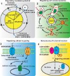
          Vitamin D (VD) reduces the risk of breast cancer and improves disease prognoses. Potential VD analogs are being developed as therapeutic agents for breast cancer treatments. The large-conductance Ca 2+-activated K + channel K Ca1.1 regulates intracellular Ca 2+ signaling pathways and is associated with high grade tumors and poor prognoses. In the present study, we examined the effects of treatments with VD receptor (VDR) agonists on the expression and activity of K Ca1.1 in human breast cancer MDA-MB-453 cells using real-time PCR, Western blotting, flow cytometry, and voltage-sensitive dye imaging. Treatments with VDR agonists for 72 h markedly decreased the expression levels of K Ca1.1 transcripts and proteins in MDA-MB-453 cells, resulting in the significant inhibition of depolarization responses induced by paxilline, a specific K Ca1.1 blocker. The specific proteasome inhibitor MG132 suppressed VDR agonist-induced decreases in K Ca1.1 protein expression. These results suggest that K Ca1.1 is a new downstream target of VDR signaling and the down-regulation of K Ca1.1 through the transcriptional repression of K Ca1.1 and enhancement of K Ca1.1 protein degradation contribute, at least partly, to the antiproliferative effects of VDR agonists in breast cancer cells.

          Related collections

          Most cited references51

          • Record: found
          • Abstract: found
          • Article: found
          Is Open Access

          Targeting potassium channels in cancer

          Potassium channels are pore-forming transmembrane proteins that regulate a multitude of biological processes by controlling potassium flow across cell membranes. Aberrant potassium channel functions contribute to diseases such as epilepsy, cardiac arrhythmia, and neuromuscular symptoms collectively known as channelopathies. Increasing evidence suggests that cancer constitutes another category of channelopathies associated with dysregulated channel expression. Indeed, potassium channel–modulating agents have demonstrated antitumor efficacy. Potassium channels regulate cancer cell behaviors such as proliferation and migration through both canonical ion permeation–dependent and noncanonical ion permeation–independent functions. Given their cell surface localization and well-known pharmacology, pharmacological strategies to target potassium channel could prove to be promising cancer therapeutics.
            Bookmark
            • Record: found
            • Abstract: found
            • Article: found
            Is Open Access

            Vitamin D and the epigenome

            Epigenetic mechanisms play a crucial role in regulating gene expression. The main mechanisms involve methylation of DNA and covalent modifications of histones by methylation, acetylation, phosphorylation, or ubiquitination. The complex interplay of different epigenetic mechanisms is mediated by enzymes acting in the nucleus. Modifications in DNA methylation are performed mainly by DNA methyltransferases (DNMTs) and ten-eleven translocation (TET) proteins, while a plethora of enzymes, such as histone acetyltransferases (HATs), histone deacetylases (HDACs), histone methyltransferases (HMTs), and histone demethylases (HDMs) regulate covalent histone modifications. In many diseases, such as cancer, the epigenetic regulatory system is often disturbed. Vitamin D interacts with the epigenome on multiple levels. Firstly, critical genes in the vitamin D signaling system, such as those coding for vitamin D receptor (VDR) and the enzymes 25-hydroxylase (CYP2R1), 1α-hydroxylase (CYP27B1), and 24-hydroxylase (CYP24A1) have large CpG islands in their promoter regions and therefore can be silenced by DNA methylation. Secondly, VDR protein physically interacts with coactivator and corepressor proteins, which in turn are in contact with chromatin modifiers, such as HATs, HDACs, HMTs, and with chromatin remodelers. Thirdly, a number of genes encoding for chromatin modifiers and remodelers, such as HDMs of the Jumonji C (JmjC)-domain containing proteins and lysine-specific demethylase (LSD) families are primary targets of VDR and its ligands. Finally, there is evidence that certain VDR ligands have DNA demethylating effects. In this review we will discuss regulation of the vitamin D system by epigenetic modifications and how vitamin D contributes to the maintenance of the epigenome, and evaluate its impact in health and disease.
              Bookmark
              • Record: found
              • Abstract: found
              • Article: not found

              Serum protein binding of 1,25-dihydroxyvitamin D: a reevaluation by direct measurement of free metabolite levels.

              Using the technique of centrifugal ultrafiltration isodialysis to measure the free concentration of 1,25-dihydroxyvitamin D [1,25-(OH)2D], we determined the affinity of serum proteins for 1,25-(OH)2D both by Scatchard analysis (increasing ligand concentration at fixed binding site concentrations) and by a novel analysis in which the binding site concentrations were varied (serial dilution) at fixed ligand concentrations. The high affinity binding constant in serum for 1,25-(OH)2D was 3.7 X 10(7) M-1 by Scatchard analysis and 4.2 X 10(7) M-1 by serial dilution analysis. Human serum albumin had a much lower affinity for 1,25-(OH)2D (5.4 X 10(4) M-1). When vitamin D-binding protein (DBP) was selectively removed from serum by an actin affinity column, the affinity of the remaining serum proteins for 1,25-(OH)2D was that of albumin. Postulating a two-site model (DBP and albumin) for transport of 1,25-(OH)2D in serum and incorporating the estimated affinity constants of DBP and albumin for this metabolite, we calculated that 85% of total circulating 1,25-(OH)2D is transported in blood bound to DBP in normal individuals (0.4% is free and 14.6% is bound to albumin). In patients with liver disease, 73% is bound to DBP (1.1% is free and 25.9% is bound to albumin). Using this same two site model, we found a reasonable correlation (r = 0.612; P less than 0.001) between the measured free 1,25-(OH)2D level and the calculated free 1,25-(OH)2D level in serum based on albumin and DBP concentrations in 16 normal subjects and 16 patients with liver disease. These results confirm the concept that although DBP is the principal protein carrier of 1,25-(OH)2D in serum, albumin is a major secondary carrier, especially in patients with low DBP levels.
                Bookmark

                Author and article information

                Contributors
                Role: Academic Editor
                Journal
                Int J Mol Sci
                Int J Mol Sci
                ijms
                International Journal of Molecular Sciences
                MDPI
                1422-0067
                11 December 2016
                December 2016
                : 17
                : 12
                : 2083
                Affiliations
                [1 ]Department of Pharmacology, Division of Pathological Sciences, Kyoto Pharmaceutical University, Kyoto 607-8414, Japan; anowarakhatun07@ 123456gmail.com (A.K.); ky13291@ 123456poppy.kyoto-phu.ac.jp (M.F.); kito@ 123456mb.kyoto-phu.ac.jp (H.K.); sniwa@ 123456mb.kyoto-phu.ac.jp (S.N.)
                [2 ]Graduate School of Medical Science, Kyoto Prefectural University of Medicine, Kyoto 403-8334, Japan; suzukit@ 123456koto.kpu-m.ac.jp
                Author notes
                [* ]Correspondence: sohya@ 123456mb.kyoto-phu.ac.jp ; Tel.: +81-75-595-4667
                Article
                ijms-17-02083
                10.3390/ijms17122083
                5187883
                27973439
                38ff72e7-f875-428f-bcaf-35b39aba2f45
                © 2016 by the authors; licensee MDPI, Basel, Switzerland.

                This article is an open access article distributed under the terms and conditions of the Creative Commons Attribution (CC-BY) license ( http://creativecommons.org/licenses/by/4.0/).

                History
                : 27 September 2016
                : 08 December 2016
                Categories
                Article

                Molecular biology
                vitamin d receptor,breast cancer,kca1.1,k+ channel,transcription,protein degradation
                Molecular biology
                vitamin d receptor, breast cancer, kca1.1, k+ channel, transcription, protein degradation

                Comments

                Comment on this article