29
views
0
recommends
+1 Recommend
0 collections
    0
    shares
      • Record: found
      • Abstract: found
      • Article: found
      Is Open Access

      Improving CT-guided transthoracic biopsy of mediastinal lesions by diffusion-weighted magnetic resonance imaging

      research-article

      Read this article at

      Bookmark
          There is no author summary for this article yet. Authors can add summaries to their articles on ScienceOpen to make them more accessible to a non-specialist audience.

          Abstract

          OBJECTIVES:

          To evaluate the preliminary results obtained using diffusion-weighted magnetic resonance imaging and the apparent diffusion coefficient for planning computed tomography-guided biopsies of selected mediastinal lesions.

          METHODS:

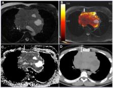
          Eight patients with mediastinal lesions suspicious for malignancy were referred for computed tomography-guided biopsy. Diffusion-weighted magnetic resonance imaging and apparent diffusion coefficient measurement were performed to assist in biopsy planning with diffusion/computed tomography fused images. We selected mediastinal lesions that could provide discordant diagnoses depending on the biopsy site, including large heterogeneous masses, lesions associated with lung atelectasis or consolidation, lesions involving large mediastinal vessels and lesions for which the results of biopsy using other methods and histopathological examination were divergent from the clinical and radiological suspicion.

          RESULTS:

          In all cases, the biopsy needle was successfully directed to areas of higher signal intensity on diffusion-weighted sequences and the lowest apparent diffusion coefficient within the lesion (mean, 0.8 [range, 0.6–1.1]×10 -3 mm 2/s), suggesting high cellularity. All biopsies provided adequate material for specific histopathological diagnoses of four lymphomas, two sarcomas and two thymomas.

          CONCLUSION:

          Functional imaging tools, such as diffusion-weighted imaging and the apparent diffusion coefficient, are promising for implementation in noninvasive and imaging-guided procedures. However, additional studies are needed to confirm that mediastinal biopsy can be improved with these techniques.

          Related collections

          Most cited references72

          • Record: found
          • Abstract: found
          • Article: not found

          Radiation exposure from CT scans in childhood and subsequent risk of leukaemia and brain tumours: a retrospective cohort study

          Summary Background Although CT scans are very useful clinically, potential cancer risks exist from associated ionising radiation, in particular for children who are more radiosensitive than adults. We aimed to assess the excess risk of leukaemia and brain tumours after CT scans in a cohort of children and young adults. Methods In our retrospective cohort study, we included patients without previous cancer diagnoses who were first examined with CT in National Health Service (NHS) centres in England, Wales, or Scotland (Great Britain) between 1985 and 2002, when they were younger than 22 years of age. We obtained data for cancer incidence, mortality, and loss to follow-up from the NHS Central Registry from Jan 1, 1985, to Dec 31, 2008. We estimated absorbed brain and red bone marrow doses per CT scan in mGy and assessed excess incidence of leukaemia and brain tumours cancer with Poisson relative risk models. To avoid inclusion of CT scans related to cancer diagnosis, follow-up for leukaemia began 2 years after the first CT and for brain tumours 5 years after the first CT. Findings During follow-up, 74 of 178 604 patients were diagnosed with leukaemia and 135 of 176 587 patients were diagnosed with brain tumours. We noted a positive association between radiation dose from CT scans and leukaemia (excess relative risk [ERR] per mGy 0·036, 95% CI 0·005–0·120; p=0·0097) and brain tumours (0·023, 0·010–0·049; p<0·0001). Compared with patients who received a dose of less than 5 mGy, the relative risk of leukaemia for patients who received a cumulative dose of at least 30 mGy (mean dose 51·13 mGy) was 3·18 (95% CI 1·46–6·94) and the relative risk of brain cancer for patients who received a cumulative dose of 50–74 mGy (mean dose 60·42 mGy) was 2·82 (1·33–6·03). Interpretation Use of CT scans in children to deliver cumulative doses of about 50 mGy might almost triple the risk of leukaemia and doses of about 60 mGy might triple the risk of brain cancer. Because these cancers are relatively rare, the cumulative absolute risks are small: in the 10 years after the first scan for patients younger than 10 years, one excess case of leukaemia and one excess case of brain tumour per 10 000 head CT scans is estimated to occur. Nevertheless, although clinical benefits should outweigh the small absolute risks, radiation doses from CT scans ought to be kept as low as possible and alternative procedures, which do not involve ionising radiation, should be considered if appropriate. Funding US National Cancer Institute and UK Department of Health.
            Bookmark
            • Record: found
            • Abstract: found
            • Article: not found

            Diffusion-weighted magnetic resonance imaging as a cancer biomarker: consensus and recommendations.

            On May 3, 2008, a National Cancer Institute (NCI)-sponsored open consensus conference was held in Toronto, Ontario, Canada, during the 2008 International Society for Magnetic Resonance in Medicine Meeting. Approximately 100 experts and stakeholders summarized the current understanding of diffusion-weighted magnetic resonance imaging (DW-MRI) and reached consensus on the use of DW-MRI as a cancer imaging biomarker. DW-MRI should be tested as an imaging biomarker in the context of well-defined clinical trials, by adding DW-MRI to existing NCI-sponsored trials, particularly those with tissue sampling or survival indicators. Where possible, DW-MRI measurements should be compared with histologic indices including cellularity and tissue response. There is a need for tissue equivalent diffusivity phantoms; meanwhile, simple fluid-filled phantoms should be used. Monoexponential assessments of apparent diffusion coefficient values should use two b values (>100 and between 500 and 1000 mm2/sec depending on the application). Free breathing with multiple acquisitions is superior to complex gating techniques. Baseline patient reproducibility studies should be part of study designs. Both region of interest and histogram analysis of apparent diffusion coefficient measurements should be obtained. Standards for measurement, analysis, and display are needed. Annotated data from validation studies (along with outcome measures) should be made publicly available. Magnetic resonance imaging vendors should be engaged in this process. The NCI should establish a task force of experts (physicists, radiologists, and oncologists) to plan, organize technical aspects, and conduct pilot trials. The American College of Radiology Imaging Network infrastructure may be suitable for these purposes. There is an extraordinary opportunity for DW-MRI to evolve into a clinically valuable imaging tool, potentially important for drug development.
              Bookmark
              • Record: found
              • Abstract: found
              • Article: not found

              Thymic epithelial tumors: comparison of CT and MR imaging findings of low-risk thymomas, high-risk thymomas, and thymic carcinomas.

              To assess the CT and magnetic resonance (MR) imaging findings of thymic epithelial tumors classified according to the current World Health Organization (WHO) histologic classification and to determine useful findings in differentiating the main subtypes. Sixty patients with thymic epithelial tumor who underwent both CT and MR imaging were reviewed retrospectively. All cases were classified according to the 2004 WHO classification. The following findings were assessed in each case on both CT and MRI: size of tumor, contour, perimeter of capsule; homogeneity, presence of septum, hemorrhage, necrotic or cystic component within tumor; presence of mediastinal lymphadenopathy, pleural effusion, and great vessel invasion. These imaging characteristics of 30 low-risk thymomas (4 type A, 12 type AB, and 14 type B1), 18 high-risk thymomas (11 type B2 and seven type B3), and 12 thymic carcinomas on CT and MR imaging were compared using the chi-square test. Comparison between CT and MR findings was performed by using McNemar test. On both CT and MR imaging, thymic carcinomas were more likely to have irregular contours (P < .001), necrotic or cystic component (P < .05), heterogeneous contrast-enhancement (P < .05), lymphadenopathy (P < .0001), and great vessel invasion (P < .001) than low-risk and high-risk thymomas. On MR imaging, the findings of almost complete capsule, septum, and homogenous enhancement were more commonly seen in low-risk thymomas than high-risk thymomas and thymic carcinomas (P < .05). MR imaging was superior to CT in the depiction of capsule, septum, or hemorrhage within tumor (all comparison, P < .05). The presence of irregular contour, necrotic or cystic component, heterogeneous enhancement, lymphadenopathy, and great vessel invasion on CT or MR imaging are strongly suggestive of thymic carcinomas. On MR imaging, the findings of contour, capsule, septum, and homogenous enhancement are helpful in distinguishing low-risk thymomas from high-risk thymomas and thymic carcinomas.
                Bookmark

                Author and article information

                Journal
                Clinics (Sao Paulo)
                Clinics (Sao Paulo)
                Clinics
                Hospital das Clínicas da Faculdade de Medicina da Universidade de São Paulo
                1807-5932
                1980-5322
                November 2014
                : 69
                : 11
                : 787-791
                Affiliations
                [I ]AC Camargo Cancer Center, Department of Imaging, São Paulo/SP, Brazil.
                [II ]Universidade Federal de Ciências da Saúde de Porto Alegre, Department of Radiology, Porto Alegre/RS, Brazil.
                [III ]University of Texas, MD Anderson Cancer Center, Department of Diagnostic Radiology, Houston, TX, USA.
                [IV ]University of Texas, MD Anderson Cancer Center, Department of Diagnostic and Interventional Radiology, Houston, TX, USA.
                [V ]AC Camargo Cancer Center, Department of Thoracic Surgery, São Paulo/SP, Brazil.
                [VI ]AC Camargo Cancer Center, Department of Interventional Radiology, São Paulo/SP, Brazil.
                [VII ]Universidade Federal do Rio de Janeiro, Department of Radiology, Petrópolis/RJ, Brazil.
                Author notes

                Guimaraes MD was the principal investigator and takes public responsibility for the entire manuscript, contributed to the coordination and study design, statistics, data interpretation and final review of the manuscript. Hochhegger B contributed to the study design, statistics, data interpretation, literature review and revision of the manuscript. Benveniste MF contributed to study design, data interpretation, literature review and revision of the manuscript. Odisio BC contributed to data collection, CT high-resolution scan evaluation, literature review, data interpretation and preparation of the manuscript. Gross JL contributed to the data collection, literature review and preparation of the manuscript. Zurstrassen E contributed to data collection, MR high-resolution evaluation, literature review and preparation of the manuscript. Tyng CC contributed to data collection, MR evaluation, literature review and preparation of the manuscript. Bitencourt AG contributed to data collection, literature review and preparation of the manuscript. Marchiori E contributed to study design, data interpretation and final review of the manuscript.

                *corresponding author
                E-mail: marcosduarte500@ 123456gmail.com Tel.: 55 11 3208-5327
                Article
                cln_69p787
                10.6061/clinics/2014(11)13
                4255081
                25518038
                3a0215e7-908b-4df6-9ade-84bfcb8b3c68
                Copyright © 2014 Hospital das Clínicas da FMUSP

                This is an Open Access article distributed under the terms of the Creative Commons Attribution Non-Commercial License ( http://creativecommons.org/licenses/by-nc/3.0/) which permits unrestricted non-commercial use, distribution, and reproduction in any medium, provided the original work is properly cited.

                History
                : 10 May 2014
                : 25 June 2014
                : 17 July 2014
                Page count
                Pages: 5
                Categories
                Rapid Communication

                Medicine
                mri-guided biopsy,dwi-guided biopsy,mediastinal lesion,diagnosis,malignancy,tomography
                Medicine
                mri-guided biopsy, dwi-guided biopsy, mediastinal lesion, diagnosis, malignancy, tomography

                Comments

                Comment on this article