1
views
0
recommends
+1 Recommend
0 collections
    0
    shares
      • Record: found
      • Abstract: found
      • Article: found
      Is Open Access

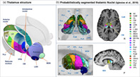
      Probabilistic mapping of thalamic nuclei and thalamocortical functional connectivity in idiopathic generalised epilepsy

      research-article

      Read this article at

      Bookmark
          There is no author summary for this article yet. Authors can add summaries to their articles on ScienceOpen to make them more accessible to a non-specialist audience.

          Abstract

          It is well established that abnormal thalamocortical systems play an important role in the generation and maintenance of primary generalised seizures. However, it is currently unknown which thalamic nuclei and how nuclear‐specific thalamocortical functional connectivity are differentially impacted in patients with medically refractory and non‐refractory idiopathic generalised epilepsy (IGE). In the present study, we performed structural and resting‐state functional magnetic resonance imaging (MRI) in patients with refractory and non‐refractory IGE, segmented the thalamus into constituent nuclear regions using a probabilistic MRI segmentation method and determined thalamocortical functional connectivity using seed‐to‐voxel connectivity analyses. We report significant volume reduction of the left and right anterior thalamic nuclei only in patients with refractory IGE. Compared to healthy controls, patients with refractory and non‐refractory IGE had significant alterations of functional connectivity between the centromedian nucleus and cortex, but only patients with refractory IGE had altered cortical connectivity with the ventral lateral nuclear group. Patients with refractory IGE had significantly increased functional connectivity between the left and right ventral lateral posterior nuclei and cortical regions compared to patients with non‐refractory IGE. Cortical effects were predominantly located in the frontal lobe. Atrophy of the anterior thalamic nuclei and resting‐state functional hyperconnectivity between ventral lateral nuclei and cerebral cortex may be imaging markers of pharmacoresistance in patients with IGE. These structural and functional abnormalities fit well with the known importance of thalamocortical systems in the generation and maintenance of primary generalised seizures, and the increasing recognition of the importance of limbic pathways in IGE.

          Abstract

          We report that patients with idiopathic generalised epilepsy have different patterns of thalamic structural and functional connectivity alterations depending on whether they are refractory or not to anti‐seizure medication. Only refractory patients showed evidence of atrophy of the anterior (limbic) thalamic nuclei and increased functional connectivity between the ventral lateral posterior nuclei and cortex bilaterally.

          Related collections

          Most cited references141

          • Record: found
          • Abstract: not found
          • Article: not found

          Controlling the False Discovery Rate: A Practical and Powerful Approach to Multiple Testing

            Bookmark
            • Record: found
            • Abstract: found
            • Article: not found

            FreeSurfer.

            FreeSurfer is a suite of tools for the analysis of neuroimaging data that provides an array of algorithms to quantify the functional, connectional and structural properties of the human brain. It has evolved from a package primarily aimed at generating surface representations of the cerebral cortex into one that automatically creates models of most macroscopically visible structures in the human brain given any reasonable T1-weighted input image. It is freely available, runs on a wide variety of hardware and software platforms, and is open source. Copyright © 2012 Elsevier Inc. All rights reserved.
              Bookmark
              • Record: found
              • Abstract: found
              • Article: not found

              Conn: a functional connectivity toolbox for correlated and anticorrelated brain networks.

              Resting state functional connectivity reveals intrinsic, spontaneous networks that elucidate the functional architecture of the human brain. However, valid statistical analysis used to identify such networks must address sources of noise in order to avoid possible confounds such as spurious correlations based on non-neuronal sources. We have developed a functional connectivity toolbox Conn ( www.nitrc.org/projects/conn ) that implements the component-based noise correction method (CompCor) strategy for physiological and other noise source reduction, additional removal of movement, and temporal covariates, temporal filtering and windowing of the residual blood oxygen level-dependent (BOLD) contrast signal, first-level estimation of multiple standard functional connectivity magnetic resonance imaging (fcMRI) measures, and second-level random-effect analysis for resting state as well as task-related data. Compared to methods that rely on global signal regression, the CompCor noise reduction method allows for interpretation of anticorrelations as there is no regression of the global signal. The toolbox implements fcMRI measures, such as estimation of seed-to-voxel and region of interest (ROI)-to-ROI functional correlations, as well as semipartial correlation and bivariate/multivariate regression analysis for multiple ROI sources, graph theoretical analysis, and novel voxel-to-voxel analysis of functional connectivity. We describe the methods implemented in the Conn toolbox for the analysis of fcMRI data, together with examples of use and interscan reliability estimates of all the implemented fcMRI measures. The results indicate that the CompCor method increases the sensitivity and selectivity of fcMRI analysis, and show a high degree of interscan reliability for many fcMRI measures.
                Bookmark

                Author and article information

                Contributors
                simon.keller@liverpool.ac.uk
                Journal
                Hum Brain Mapp
                Hum Brain Mapp
                10.1002/(ISSN)1097-0193
                HBM
                Human Brain Mapping
                John Wiley & Sons, Inc. (Hoboken, USA )
                1065-9471
                1097-0193
                25 August 2021
                1 December 2021
                : 42
                : 17 ( doiID: 10.1002/hbm.v42.17 )
                : 5648-5664
                Affiliations
                [ 1 ] Department of Pharmacology and Therapeutics Institute of Systems, Molecular and Integrative Biology, University of Liverpool Liverpool UK
                [ 2 ] The Walton Centre NHS Foundation Trust Liverpool UK
                [ 3 ] Department of Psychology University of Liverpool Liverpool UK
                [ 4 ] Department of Neurology University Medicine Göttingen Göttingen Germany
                [ 5 ] Schools of Medical Sciences and Psychology, Bangor University Bangor UK
                [ 6 ] Department of Neurology Manchester Centre for Clinical Neurosciences, Salford Royal NHS Foundation Trust Salford UK
                [ 7 ] Division of Neuroscience and Experimental Psychology, Faculty of Biology, Medicine and Health School of Biological Sciences, University of Manchester Manchester UK
                Author notes
                [*] [* ] Correspondence

                Simon S. Keller Clinical Sciences Centre, Aintree University Hospital, Lower Lane, Liverpool, L9 7LJ, UK.

                Email: simon.keller@ 123456liverpool.ac.uk

                Author information
                https://orcid.org/0000-0001-5247-9795
                Article
                HBM25644
                10.1002/hbm.25644
                8559489
                34432348
                3ce058a6-a73a-40ae-82f5-375dc8f583df
                © 2021 The Authors. Human Brain Mapping published by Wiley Periodicals LLC.

                This is an open access article under the terms of the http://creativecommons.org/licenses/by/4.0/ License, which permits use, distribution and reproduction in any medium, provided the original work is properly cited.

                History
                : 04 August 2021
                : 01 May 2021
                : 16 August 2021
                Page count
                Figures: 5, Tables: 5, Pages: 17, Words: 15066
                Funding
                Funded by: Epilepsy Research UK , doi 10.13039/501100000295;
                Award ID: 1085
                Funded by: Medical Research Council , doi 10.13039/501100000265;
                Award ID: MR/S00355X/1
                Award ID: MR/K023152/1
                Categories
                Research Article
                Research Articles
                Custom metadata
                2.0
                December 1, 2021
                Converter:WILEY_ML3GV2_TO_JATSPMC version:6.0.8 mode:remove_FC converted:01.11.2021

                Neurology
                epilepsy,functional mri,pharmacoresistance,thalamus
                Neurology
                epilepsy, functional mri, pharmacoresistance, thalamus

                Comments

                Comment on this article