9
views
0
recommends
+1 Recommend
0 collections
    0
    shares
      • Record: found
      • Abstract: found
      • Article: found
      Is Open Access

      Autophagy enhances the replication of Peste des petits ruminants virus and inhibits caspase-dependent apoptosis in vitro

      research-article

      Read this article at

      Bookmark
          There is no author summary for this article yet. Authors can add summaries to their articles on ScienceOpen to make them more accessible to a non-specialist audience.

          ABSTRACT

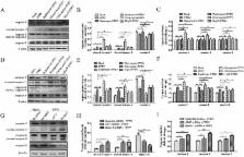
          Peste des petits ruminants (PPR) is an acute and highly contagious disease in small ruminants that causes significant economic losses in developing countries. An increasing number of studies have demonstrated that both autophagy and apoptosis are important cellular mechanisms for maintaining homeostasis, and they participate in the host response to pathogens. However, the crosstalk between apoptosis and autophagy in host cells during PPRV infection has not been clarified. In this study, autophagy was induced upon virus infection in caprine endometrial epithelial cells (EECs), as determined by the appearance of double- and single-membrane autophagy-like vesicles, LC3-I/LC3-II conversion, and p62 degradation. We also found that PPRV infection triggered a complete autophagic response, most likely mediated by the non-structural protein C and nucleoprotein N. Moreover, our results suggest that autophagy not only promotes the replication of PPRV in EECs but also provides a potential mechanism for inhibiting PPRV-induced apoptosis. Inhibiting autophagosome formation by wortmannin and knocking down the essential autophagic proteins Beclin-1 and ATG7 induces caspase-dependent apoptosis in EECs in PPRV infection. However, inhibiting autophagosome and lysosome fusion by NH 4Cl and chloroquine did not increase the number of apoptotic cells. Collectively, these data are the first to indicate that PPRV-induced autophagy inhibits caspase-dependent apoptosis and thus contributes to the enhancement of viral replication and maturity in host cells.

          Related collections

          Most cited references59

          • Record: found
          • Abstract: found
          • Article: not found

          Cell death: critical control points.

          Programmed cell death is a distinct genetic and biochemical pathway essential to metazoans. An intact death pathway is required for successful embryonic development and the maintenance of normal tissue homeostasis. Apoptosis has proven to be tightly interwoven with other essential cell pathways. The identification of critical control points in the cell death pathway has yielded fundamental insights for basic biology, as well as provided rational targets for new therapeutics.
            Bookmark
            • Record: found
            • Abstract: found
            • Article: not found

            Life and death partners: apoptosis, autophagy and the cross-talk between them.

            It is not surprising that the demise of a cell is a complex well-controlled process. Apoptosis, the first genetically programmed death process identified, has been extensively studied and its contribution to the pathogenesis of disease well documented. Yet, apoptosis does not function alone to determine a cell's fate. More recently, autophagy, a process in which de novo-formed membrane-enclosed vesicles engulf and consume cellular components, has been shown to engage in a complex interplay with apoptosis. In some cellular settings, it can serve as a cell survival pathway, suppressing apoptosis, and in others, it can lead to death itself, either in collaboration with apoptosis or as a back-up mechanism when the former is defective. The molecular regulators of both pathways are inter-connected; numerous death stimuli are capable of activating either pathway, and both pathways share several genes that are critical for their respective execution. The cross-talk between apoptosis and autophagy is therefore quite complex, and sometimes contradictory, but surely critical to the overall fate of the cell. Furthermore, the cross-talk is a key factor in the outcome of death-related pathologies such as cancer, its development and treatment.
              Bookmark
              • Record: found
              • Abstract: found
              • Article: not found

              How to interpret LC3 immunoblotting.

              Microtubule-associated protein light chain 3 (LC3) is now widely used to monitor autophagy. One approach is to detect LC3 conversion (LC3-I to LC3-II) by immunoblot analysis because the amount of LC3-II is clearly correlated with the number of autophagosomes. However, LC3-II itself is degraded by autophagy, making interpretation of the results of LC3 immunoblotting problematic. Furthermore, the amount of LC3 at a certain time point does not indicate autophagic flux, and therefore, it is important to measure the amount of LC3-II delivered to lysosomes by comparing LC3-II levels in the presence and absence of lysosomal protease inhibitors. Another problem with this method is that LC3-II tends to be much more sensitive to be detected by immunoblotting than LC3-I. Accordingly, simple comparison of LC3-I and LC3-II, or summation of LC3-I and LC3-II for ratio determinations, may not be appropriate, and rather, the amount of LC3-II can be compared between samples.
                Bookmark

                Author and article information

                Journal
                Virulence
                Virulence
                KVIR
                kvir20
                Virulence
                Taylor & Francis
                2150-5594
                2150-5608
                2018
                1 August 2018
                1 August 2018
                : 9
                : 1
                : 1176-1194
                Affiliations
                [a ]College of Veterinary Medicine, Northwest A&F University , Yangling, China
                [b ]China Institute of Veterinary Drug Control , Beijing, China
                Author notes
                CONTACT Jingyu Wang nwsuaf4409@ 123456126.com College of Veterinary Medicine, Northwest A&F University , Xinong Road 22#, Yangling 712100, Shaanxi, China
                [*]

                These authors contributed equally to this work.

                Article
                1496776
                10.1080/21505594.2018.1496776
                6086290
                30067475
                3cf996da-15cb-478c-ae69-85d559e361ad
                © 2018 The Author(s). Published by Informa UK Limited, trading as Taylor & Francis Group.

                This is an Open Access article distributed under the terms of the Creative Commons Attribution License ( http://creativecommons.org/licenses/by/4.0/), which permits unrestricted use, distribution, and reproduction in any medium, provided the original work is properly cited.

                History
                : 2 February 2018
                : 25 June 2018
                Page count
                Figures: 10, References: 75, Pages: 19
                Funding
                Funded by: National Natural Science Foundation of China 10.13039/501100001809
                Award ID: 31602035
                Funded by: National Key Research and Development Program of China
                Award ID: 2017YFD0500902
                This work was supported by grants from the National Natural Science Foundation of China (31602035) and the National Key Research and Development Program of China (2017YFD0500902).
                Categories
                Research Paper

                Infectious disease & Microbiology
                peste des petits ruminants virus (pprv),autophagy,apoptosis,replication,caprine endometrial epithelial cells (eecs)

                Comments

                Comment on this article