2
views
0
recommends
+1 Recommend
0 collections
    0
    shares
      • Record: found
      • Abstract: found
      • Article: not found

      The role of activin: the other side of chronic kidney disease–mineral bone disorder?

      Read this article at

      ScienceOpenPublisherPubMed
      Bookmark
          There is no author summary for this article yet. Authors can add summaries to their articles on ScienceOpen to make them more accessible to a non-specialist audience.

          Abstract

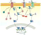
          Chronic kidney disease–mineral bone disorder (CKD-MBD) plays a pivotal role in the excess of cardiovascular morbidity and mortality associated with CKD. There is now a growing awareness that pathways involved in CKD-MBD, like canonical Wnt signalling, are activated from the earliest stages of CKD, playing a role in the development of adynamic bone disease with unknown consequences on vasculature. These changes occur before the classic changes in mineral metabolism: secondary hyperparathyroidism, calcitriol deficiency and hyperphosphataemia. Furthermore, vascular calcification is frequently associated and evolves with decreased bone mineral density and deranged bone turnover, while bone and arterial mineralization share common pathways. Therefore, results of clinical trials focused on mineral bone disorder, aimed at preserving bone and cardiovascular health, are considered unsatisfactory. In order to identify more effective therapeutic strategies, it is necessary to clarify the pathways modulating the cross-talk between bone and vasculature and identify new mediators involved in the pathogenesis of CKD-MBD. Much attention has been paid recently to the role of the transforming growth factor-beta superfamily members in renal disease, and in particular of activin A (ActA). Preclinical studies demonstrate an upgrade of ActA signalling in kidney, skeleton, vasculature and heart during CKD. This supports the idea that an endocrine factor produced in the kidney during renal disease, in addition to promoting the progression of kidney damage, deranges other organs’ homoeostasis and participates in CKD-MBD. In this review, we analyse the contribution of ActA to kidney fibrosis and inflammation as well as its role in the development of CKD-MBD.

          Related collections

          Most cited references53

          • Record: found
          • Abstract: found
          • Article: not found

          FGF23 induces left ventricular hypertrophy.

          Chronic kidney disease (CKD) is a public health epidemic that increases risk of death due to cardiovascular disease. Left ventricular hypertrophy (LVH) is an important mechanism of cardiovascular disease in individuals with CKD. Elevated levels of FGF23 have been linked to greater risks of LVH and mortality in patients with CKD, but whether these risks represent causal effects of FGF23 is unknown. Here, we report that elevated FGF23 levels are independently associated with LVH in a large, racially diverse CKD cohort. FGF23 caused pathological hypertrophy of isolated rat cardiomyocytes via FGF receptor-dependent activation of the calcineurin-NFAT signaling pathway, but this effect was independent of klotho, the coreceptor for FGF23 in the kidney and parathyroid glands. Intramyocardial or intravenous injection of FGF23 in wild-type mice resulted in LVH, and klotho-deficient mice demonstrated elevated FGF23 levels and LVH. In an established animal model of CKD, treatment with an FGF-receptor blocker attenuated LVH, although no change in blood pressure was observed. These results unveil a klotho-independent, causal role for FGF23 in the pathogenesis of LVH and suggest that chronically elevated FGF23 levels contribute directly to high rates of LVH and mortality in individuals with CKD.
            Bookmark
            • Record: found
            • Abstract: found
            • Article: not found

            Growth differentiation factor 11 is a circulating factor that reverses age-related cardiac hypertrophy.

            The most common form of heart failure occurs with normal systolic function and often involves cardiac hypertrophy in the elderly. To clarify the biological mechanisms that drive cardiac hypertrophy in aging, we tested the influence of circulating factors using heterochronic parabiosis, a surgical technique in which joining of animals of different ages leads to a shared circulation. After 4 weeks of exposure to the circulation of young mice, cardiac hypertrophy in old mice dramatically regressed, accompanied by reduced cardiomyocyte size and molecular remodeling. Reversal of age-related hypertrophy was not attributable to hemodynamic or behavioral effects of parabiosis, implicating a blood-borne factor. Using modified aptamer-based proteomics, we identified the TGF-β superfamily member GDF11 as a circulating factor in young mice that declines with age. Treatment of old mice to restore GDF11 to youthful levels recapitulated the effects of parabiosis and reversed age-related hypertrophy, revealing a therapeutic opportunity for cardiac aging. Copyright © 2013 Elsevier Inc. All rights reserved.
              Bookmark
              • Record: found
              • Abstract: found
              • Article: found
              Is Open Access

              The Role of the TGF-β Superfamily in Myocardial Infarction

              The members of the transforming growth factor β (TGF-β) superfamily are essential regulators of cell differentiation, phenotype and function, and have been implicated in the pathogenesis of many diseases. Myocardial infarction is associated with induction of several members of the superfamily, including TGF-β1, TGF-β2, TGF-β3, bone morphogenetic protein (BMP)-2, BMP-4, BMP-10, growth differentiation factor (GDF)-8, GDF-11 and activin A. This manuscript reviews our current knowledge on the patterns and mechanisms of regulation and activation of TGF-β superfamily members in the infarcted heart, and discusses their cellular actions and downstream signaling mechanisms. In the infarcted heart, TGF-β isoforms modulate cardiomyocyte survival and hypertrophic responses, critically regulate immune cell function, activate fibroblasts, and stimulate a matrix-preserving program. BMP subfamily members have been suggested to exert both pro- and anti-inflammatory actions and may regulate fibrosis. Members of the GDF subfamily may also modulate survival and hypertrophy of cardiomyocytes and regulate inflammation. Important actions of TGF-β superfamily members may be mediated through activation of Smad-dependent or non-Smad pathways. The critical role of TGF-β signaling cascades in cardiac repair, remodeling, fibrosis, and regeneration may suggest attractive therapeutic targets for myocardial infarction patients. However, the pleiotropic, cell-specific, and context-dependent actions of TGF-β superfamily members pose major challenges in therapeutic translation.
                Bookmark

                Author and article information

                Contributors
                (View ORCID Profile)
                Journal
                Nephrology Dialysis Transplantation
                Oxford University Press (OUP)
                0931-0509
                1460-2385
                June 01 2021
                May 27 2021
                September 17 2020
                June 01 2021
                May 27 2021
                September 17 2020
                : 36
                : 6
                : 966-974
                Affiliations
                [1 ]Department of Experimental Diagnostic and Specialty Medicine (DIMES), Nephrology, Dialysis and Renal Transplant Unit, S. Orsola Hospital, University of Bologna, Bologna, Italy
                [2 ]Department of Health Sciences, Renal Division, ASST Santi Paolo e Carlo, University of Milan, Milan, Italy
                [3 ]Department of Nephrology, Dialysis and Renal Transplant, Renal Research Laboratory, Fondazione IRCCS Ca’ Granda, Ospedale Maggiore Policlinico, Milan, Italy
                Article
                10.1093/ndt/gfaa203
                32940690
                3d796a49-b0a7-4f79-b25b-711ab0de611c
                © 2020

                https://academic.oup.com/journals/pages/open_access/funder_policies/chorus/standard_publication_model

                History

                Comments

                Comment on this article