8
views
0
recommends
+1 Recommend
0 collections
    0
    shares
      • Record: found
      • Abstract: found
      • Article: found
      Is Open Access

      STAT3 is Overactivated in Gastric Cancer Stem-Like Cells

      research-article

      Read this article at

      Bookmark
          There is no author summary for this article yet. Authors can add summaries to their articles on ScienceOpen to make them more accessible to a non-specialist audience.

          Abstract

          Objective

          Gastric cancer (GC) is widely associated with chronic inflammation. The pro inflammatory microenvironment provides conditions that disrupt stem/progenitor cell proliferation and differentiation. The signal transducer and activator of transcrip- tion-3 (STAT3) signaling pathway is involved in inflammation and also contributes to the maintenance of embryonic stem cell (ESCs) pluripotency. Here, we have investi- gated the activation status of STAT3 in GC stem-like cells (GCSLCs).

          Materials and Methods

          In this experimental research, CSLCs derived from the human GC cell line MKN-45 and patient specimens, through spheroid body formation, character- ized and then assayed for the STAT3 transcription factor expression in mRNA and protein level further to its activation.
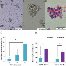
          Results

          Spheroid cells showed higher potential for spheroid formation than the pa- rental cells. Furthemore, stemness genes NANOG, c-MYC and SOX-2 were over expressed in spheroids of MKN-45 and in patient samples. In MKN-45 spheroid cells, epithelial mesenchymal transition (EMT) related markers CDH2, SNAIL2, TWIST and VIMENTIN were upregulated (P<0.05), but we observed no change in expression of the E-cadherin epithelial marker. These cells exhibited more resistance to docetaxel (DTX) when compared with parental cells (P<0.05) according to the MTS assay. Al- though immunostaining and Western blotting showed expression of the STAT3 pro- tein in both spheroids and parents, the mRNA level of STAT3 in spheroids was higher than the parents. Nuclear translocation of STAT3 was accompanied by more intensive phospho-STAT3 (p-STAT3) in spheroid structures relative to the parent cells accord- ing to flow cytometry analysis (P<0.05).

          Conclusion

          The present findings point to STAT3 over activation in GCSLCs. Com- plementary experiments are required to extend the role of STAT3 in stemness fea- tures and invasion properties of GCSCs and to consider the STAT3 pathway for CSC targeted therapy.

          Related collections

          Most cited references41

          • Record: found
          • Abstract: found
          • Article: not found

          The JAK2/STAT3 signaling pathway is required for growth of CD44⁺CD24⁻ stem cell-like breast cancer cells in human tumors.

          Intratumor heterogeneity is a major clinical problem because tumor cell subtypes display variable sensitivity to therapeutics and may play different roles in progression. We previously characterized 2 cell populations in human breast tumors with distinct properties: CD44+CD24- cells that have stem cell-like characteristics, and CD44-CD24+ cells that resemble more differentiated breast cancer cells. Here we identified 15 genes required for cell growth or proliferation in CD44+CD24- human breast cancer cells in a large-scale loss-of-function screen and found that inhibition of several of these (IL6, PTGIS, HAS1, CXCL3, and PFKFB3) reduced Stat3 activation. We found that the IL-6/JAK2/Stat3 pathway was preferentially active in CD44+CD24- breast cancer cells compared with other tumor cell types, and inhibition of JAK2 decreased their number and blocked growth of xenografts. Our results highlight the differences between distinct breast cancer cell types and identify targets such as JAK2 and Stat3 that may lead to more specific and effective breast cancer therapies.
            Bookmark
            • Record: found
            • Abstract: not found
            • Article: not found

            Schistosomes, liver flukes and Helicobacter pylori. IARC Working Group on the Evaluation of Carcinogenic Risks to Humans. Lyon, 7-14 June 1994.

              Bookmark
              • Record: found
              • Abstract: found
              • Article: not found

              Inflammation and EMT: an alliance towards organ fibrosis and cancer progression

              Recent advances in our understanding of the molecular pathways that govern the association of inflammation with organ fibrosis and cancer point to the epithelial to mesenchymal transition (EMT) as the common link in the progression of these devastating diseases. The EMT is a crucial process in the development of different tissues in the embryo and its reactivation in the adult may be regarded as a physiological attempt to control inflammatory responses and to ‘heal’ damaged tissue. However, in pathological contexts such as in tumours or during the development of organ fibrosis, this healing response adopts a sinister nature, steering these diseases towards metastasis and organ failure. Importantly, the chronic inflammatory microenvironment common to fibrotic and cancer cells emerges as a decisive factor in the induction of the pathological EMT.
                Bookmark

                Author and article information

                Journal
                Cell J
                Cell J
                Royan Institute
                Cell Journal (Yakhteh)
                Royan Institute
                2228-5806
                2228-5814
                Winter 2016
                17 January 2016
                : 17
                : 4
                : 617-628
                Affiliations
                [1 ]Department of Immunology, Faculty of Medical Sciences, Tarbiat Modares University, Tehran, Iran
                [2 ]Department of Stem Cells and Developmental Biology, Cell Sciences Research Center, Royan Institute for Stem Cell Biology and Technology, ACECR, Tehran, Iran
                [3 ]Department of Hematology, Faculty of Medical Sciences, Tarbiat Modares University, Tehran, Iran
                [4 ]Department of Molecular Medicine, Faculty of Advanced Medical Technologies, Gorgan University of Medical Sciences, Gorgan, Iran
                Author notes
                [* Corresponding Addresses: ]P.O.Box: 14117-13116Department of ImmunologyFaculty of Medical SciencesTarbiat Modares University TehranIranP.O.Box: 16635-148Department of Stem Cells and Developmental BiologyCell Sciences Research CenterRoyan Institute for Stem Cell Biology and Technology ACECRTehranIran Emails: hasan_zm@ 123456modares.ac.ir , mebrahimi@ 123456royaninstitute.org
                Article
                Cell-J-17-617
                10.22074/cellj.2016.3834
                4746412
                26862521
                3e90babd-6c72-421e-a60b-ec2c0e7eeca2
                Any use, distribution, reproduction or abstract of this publication in any medium, with the exception of commercial purposes, is permitted provided the original work is properly cited

                This is an open-access article distributed under the terms of the Creative Commons Attribution License, which permits unrestricted use, distribution, and reproduction in any medium, provided the original work is properly cited.

                History
                : 15 March 2015
                : 9 June 2015
                Categories
                Original Article
                Stem Cells
                Oncology
                Custom metadata
                Hajimoradi M, Mohammad Hassan Z, Ebrahimi M, Soleimani M, Bakhshi M, Firouzi J, Sahraneshin Samani F. STAT3 is overactivated in gastric cancer stem-like cells. Cell J. 2016; 17(4): 617-628.

                gastric cancer,cancer stem cells,spheroid,stat3,emt
                gastric cancer, cancer stem cells, spheroid, stat3, emt

                Comments

                Comment on this article