3
views
0
recommends
+1 Recommend
0 collections
    0
    shares
      • Record: found
      • Abstract: found
      • Article: found
      Is Open Access

      Border-associated macrophages promote cerebral amyloid angiopathy and cognitive impairment through vascular oxidative stress

      research-article

      Read this article at

      Bookmark
          There is no author summary for this article yet. Authors can add summaries to their articles on ScienceOpen to make them more accessible to a non-specialist audience.

          Abstract

          Background

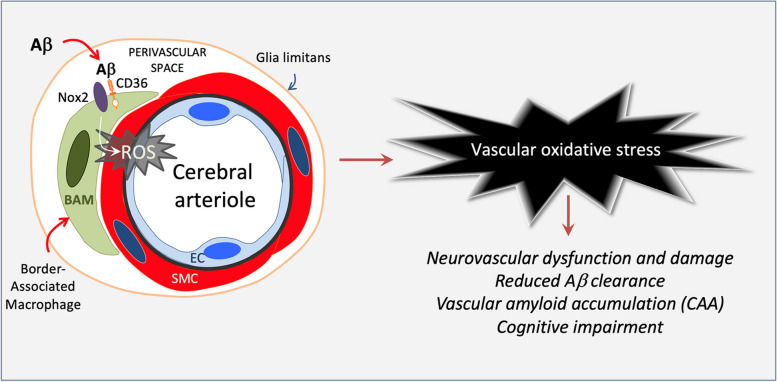
          Cerebral amyloid angiopathy (CAA) is a devastating condition common in patients with Alzheimer’s disease but also observed in the general population. Vascular oxidative stress and neurovascular dysfunction have been implicated in CAA but the cellular source of reactive oxygen species (ROS) and related signaling mechanisms remain unclear. We tested the hypothesis that brain border-associated macrophages (BAM), yolk sac-derived myeloid cells closely apposed to parenchymal and leptomeningeal blood vessels, are the source of radicals through the Aβ-binding innate immunity receptor CD36, leading to neurovascular dysfunction, CAA, and cognitive impairment.

          Methods

          Tg2576 mice and WT littermates were transplanted with CD36 −/− or CD36 +/+ bone marrow at 12-month of age and tested at 15 months. This approach enables the repopulation of perivascular and leptomeningeal compartments with CD36 −/− BAM. Neurovascular function was tested in anesthetized mice equipped with a cranial window in which cerebral blood flow was monitored by laser-Doppler flowmetry. Amyloid pathology and cognitive function were also examined.

          Results

          The increase in blood flow evoked by whisker stimulation (functional hyperemia) or by endothelial and smooth muscle vasoactivity was markedly attenuated in WT → Tg2576 chimeras but was fully restored in CD36 −/− → Tg2576 chimeras, in which BAM ROS production was suppressed. CAA-associated Aβ 1-40, but not Aβ 1-42, was reduced in CD36 −/− → Tg2576 chimeras. Similarly, CAA, but not parenchymal plaques, was reduced in CD36 −/− → Tg2576 chimeras. These beneficial vascular effects were associated with cognitive improvement. Finally, CD36 −/− mice were able to more efficiently clear exogenous Aβ 1-40 injected into the neocortex or the striatum.

          Conclusions

          CD36 deletion in BAM suppresses ROS production and rescues the neurovascular dysfunction and damage induced by Aβ. CD36 deletion in BAM also reduced brain Aβ 1-40 and ameliorated CAA without affecting parenchyma plaques. Lack of CD36 enhanced the vascular clearance of exogenous Aβ. Restoration of neurovascular function and attenuation of CAA resulted in a near complete rescue of cognitive function. Collectively, these data implicate brain BAM in the pathogenesis of CAA and raise the possibility that targeting BAM CD36 is beneficial in CAA and other conditions associated with vascular Aβ deposition and damage.

          Graphical Abstract

          Supplementary Information

          The online version contains supplementary material available at 10.1186/s13024-023-00660-1.

          Related collections

          Most cited references100

          • Record: found
          • Abstract: found
          • Article: not found

          The antibody aducanumab reduces Aβ plaques in Alzheimer's disease.

          Alzheimer's disease (AD) is characterized by deposition of amyloid-β (Aβ) plaques and neurofibrillary tangles in the brain, accompanied by synaptic dysfunction and neurodegeneration. Antibody-based immunotherapy against Aβ to trigger its clearance or mitigate its neurotoxicity has so far been unsuccessful. Here we report the generation of aducanumab, a human monoclonal antibody that selectively targets aggregated Aβ. In a transgenic mouse model of AD, aducanumab is shown to enter the brain, bind parenchymal Aβ, and reduce soluble and insoluble Aβ in a dose-dependent manner. In patients with prodromal or mild AD, one year of monthly intravenous infusions of aducanumab reduces brain Aβ in a dose- and time-dependent manner. This is accompanied by a slowing of clinical decline measured by Clinical Dementia Rating-Sum of Boxes and Mini Mental State Examination scores. The main safety and tolerability findings are amyloid-related imaging abnormalities. These results justify further development of aducanumab for the treatment of AD. Should the slowing of clinical decline be confirmed in ongoing phase 3 clinical trials, it would provide compelling support for the amyloid hypothesis.
            Bookmark
            • Record: found
            • Abstract: found
            • Article: not found

            Correlative memory deficits, Abeta elevation, and amyloid plaques in transgenic mice.

            Transgenic mice overexpressing the 695-amino acid isoform of human Alzheimer beta-amyloid (Abeta) precursor protein containing a Lys670 --> Asn, Met671 --> Leu mutation had normal learning and memory in spatial reference and alternation tasks at 3 months of age but showed impairment by 9 to 10 months of age. A fivefold increase in Abeta(1-40) and a 14-fold increase in Abeta(1-42/43) accompanied the appearance of these behavioral deficits. Numerous Abeta plaques that stained with Congo red dye were present in cortical and limbic structures of mice with elevated amounts of Abeta. The correlative appearance of behavioral, biochemical, and pathological abnormalities reminiscent of Alzheimer's disease in these transgenic mice suggests new opportunities for exploring the pathophysiology and neurobiology of this disease.
              Bookmark
              • Record: found
              • Abstract: found
              • Article: not found

              Functional aspects of meningeal lymphatics in aging and Alzheimer’s disease

              Aging is a major risk factor for many neurological pathologies and the mechanisms underlying brain aging remain elusive. Unlike other tissues, the central nervous system (CNS) parenchyma is devoid of lymphatic vasculature and removal of waste products is performed mainly through a paravascular route. (Re)discovery and characterization of meningeal lymphatic vessels prompted for an assessment of their role in CNS waste clearance. Here we show that meningeal lymphatics are draining macromolecules from the CNS (CSF and ISF) into the cervical lymph nodes. Impairment of meningeal lymphatic function slows paravascular influx of CSF macromolecules and efflux of ISF macromolecules, and induces cognitive impairment. Treatment of aged mice with vascular endothelial growth factor C enhances meningeal lymphatic drainage of CSF macromolecules, improving brain perfusion and learning and memory performance. Disruption of meningeal lymphatic vessels in transgenic mouse models of Alzheimer’s disease (AD) promotes amyloid deposition in the meninges, which closely resembles human meningeal pathology, and aggravates parenchymal amyloid accumulation. Our findings suggest that meningeal lymphatic dysfunction may be an aggravating factor in AD pathology and in age-associated cognitive decline. Thus, augmentation of meningeal lymphatic function might be a promising therapeutic target for preventing or delaying age-associated neurological diseases.
                Bookmark

                Author and article information

                Contributors
                lap2003@med.cornell.edu
                coi2001@med.cornell.edu
                Journal
                Mol Neurodegener
                Mol Neurodegener
                Molecular Neurodegeneration
                BioMed Central (London )
                1750-1326
                3 October 2023
                3 October 2023
                2023
                : 18
                : 73
                Affiliations
                Feil Family Brain and Mind Research Institute, Weill Cornell Medicine, ( https://ror.org/02r109517) New York, NY 10021 USA
                Author information
                http://orcid.org/0000-0001-9797-073X
                Article
                660
                10.1186/s13024-023-00660-1
                10548599
                37789345
                3fe63b37-39b4-48cd-b2b1-aefe24d79422
                © Editorial Group and BioMed Central Ltd., part of Springer Nature 2023

                Open Access This article is licensed under a Creative Commons Attribution 4.0 International License, which permits use, sharing, adaptation, distribution and reproduction in any medium or format, as long as you give appropriate credit to the original author(s) and the source, provide a link to the Creative Commons licence, and indicate if changes were made. The images or other third party material in this article are included in the article's Creative Commons licence, unless indicated otherwise in a credit line to the material. If material is not included in the article's Creative Commons licence and your intended use is not permitted by statutory regulation or exceeds the permitted use, you will need to obtain permission directly from the copyright holder. To view a copy of this licence, visit http://creativecommons.org/licenses/by/4.0/. The Creative Commons Public Domain Dedication waiver ( http://creativecommons.org/publicdomain/zero/1.0/) applies to the data made available in this article, unless otherwise stated in a credit line to the data.

                History
                : 27 March 2023
                : 20 September 2023
                Funding
                Funded by: FundRef http://dx.doi.org/10.13039/100000002, National Institutes of Health;
                Award ID: NS37853
                Award Recipient :
                Funded by: National Institute of Health
                Award ID: NS097805
                Award Recipient :
                Funded by: Japan Heart Foundation/Bayer Research Grant Abroad
                Funded by: The Uehara Memorial Foundation Research Fellowship
                Funded by: Japan Society for the Promotion of Science Overseas Research Fellowships
                Funded by: FundRef http://dx.doi.org/10.13039/100007027, Leon Levy Foundation;
                Funded by: FundRef http://dx.doi.org/10.13039/100006312, BrightFocus Foundation;
                Categories
                Research Article
                Custom metadata
                © Editorial Group and BioMed Central Ltd., part of Springer Nature 2023

                Neurosciences
                border-associated macrophages,cd36,aβ clearance,neurovascular unit dysfunction,vascular oxidative stress,aria

                Comments

                Comment on this article