7
views
0
recommends
+1 Recommend
0 collections
    0
    shares
      • Record: found
      • Abstract: found
      • Article: found
      Is Open Access

      Endothelial-to-mesenchymal transition in anticancer therapy and normal tissue damage

      review-article

      Read this article at

      Bookmark
          There is no author summary for this article yet. Authors can add summaries to their articles on ScienceOpen to make them more accessible to a non-specialist audience.

          Abstract

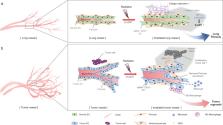
          Endothelial-to-mesenchymal transition (EndMT) involves the phenotypic conversion of endothelial-to-mesenchymal cells, and was first discovered in association with embryonic heart development. EndMT can regulate various processes, such as tissue fibrosis and cancer. Recent findings have shown that EndMT is related to resistance to cancer therapy, such as chemotherapy, antiangiogenic therapy, and radiation therapy. Based on the known effects of EndMT on the cardiac toxicity of anticancer therapy and tissue damage of radiation therapy, we propose that EndMT can be targeted as a strategy for overcoming tumor resistance while reducing complications, such as tissue damage. In this review, we discuss EndMT and its roles in damaging cardiac and lung tissues, as well as EndMT-related effects on tumor vasculature and resistance in anticancer therapy. Modulating EndMT in radioresistant tumors and radiation-induced tissue fibrosis can especially increase the efficacy of radiation therapy. In addition, we review the role of hypoxia and reactive oxygen species as the main stimulating factors of tissue damage due to vascular damage and EndMT. We consider drugs that may be clinically useful for regulating EndMT in various diseases. Finally, we argue the importance of EndMT as a therapeutic target in anticancer therapy for reducing tissue damage.

          Cell biology: effect of changes in cell type on disease

          A process of cellular conversion known as endothelial-to-mesenchymal transition (EndMT) may offer a valuable target for treating cancer and other diseases. In EndMT, the cells lining blood vessels undergo a striking change in shape and physiology, acquiring features of cells called fibroblasts. Fibroblasts form the body’s connective tissue, but also produce scar tissue that impairs organ function. Researchers led by Yoon-Jin Lee of the Korea Institute of Radiological & Medical Sciences in Seoul, South Korea, have reviewed the impact of this transformation on human disease. EndMT is seen as a prelude to heart failure, in lung tissue affected by pulmonary fibrosis, and within tumors, where the process recruits cells that further stimulate cancer progression. The authors highlight the potential of using drugs that target EndMT to bolster the efficacy and safety of tumor therapy.

          Related collections

          Most cited references90

          • Record: found
          • Abstract: found
          • Article: not found

          Kinetics of vascular normalization by VEGFR2 blockade governs brain tumor response to radiation: role of oxygenation, angiopoietin-1, and matrix metalloproteinases.

          The recent landmark Phase III clinical trial with a VEGF-specific antibody suggests that antiangiogenic therapy must be combined with cytotoxic therapy for the treatment of solid tumors. However, there are no guidelines for optimal scheduling of these therapies. Here we show that VEGFR2 blockade creates a "normalization window"--a period during which combined radiation therapy gives the best outcome. This window is characterized by an increase in tumor oxygenation, which is known to enhance radiation response. During the normalization window, but not before or after it, VEGFR2 blockade increases pericyte coverage of brain tumor vessels via upregulation of Ang1 and degrades their pathologically thick basement membrane via MMP activation.
            Bookmark
            • Record: found
            • Abstract: not found
            • Article: not found

            Cardiovascular Toxic Effects of Targeted Cancer Therapies.

              Bookmark
              • Record: found
              • Abstract: found
              • Article: not found

              Integration of TGF-beta/Smad and Jagged1/Notch signalling in epithelial-to-mesenchymal transition.

              Epithelial-to-mesenchymal transitions (EMTs) underlie cell plasticity required in embryonic development and frequently observed in advanced carcinogenesis. Transforming growth factor-beta (TGF-beta) induces EMT phenotypes in epithelial cells in vitro and has been associated with EMT in vivo. Here we report that expression of the hairy/enhancer-of-split-related transcriptional repressor Hey1, and the Notch-ligand Jagged1 (Jag1), was induced by TGF-beta at the onset of EMT in epithelial cells from mammary gland, kidney tubules, and epidermis. The HEY1 expression profile was biphasic, consisting of immediate-early Smad3-dependent, Jagged1/Notch-independent activation, followed by delayed, indirect Jagged1/Notch-dependent activation. TGF-beta-induced EMT was blocked by RNA silencing of HEY1 or JAG1, and by chemical inactivation of Notch. The EMT phenotype, biphasic activation of Hey1, and delayed expression of Jag1 were induced by TGF-beta in wild-type, but not in Smad3-deficient, primary mouse kidney tubular epithelial cells. Our findings identify a new mechanism for functional integration of Jagged1/Notch signalling and coordinated activation of the Hey1 transcriptional repressor controlled by TGF-beta/Smad3, and demonstrate functional roles for Smad3, Hey1, and Jagged1/Notch in mediating TGF-beta-induced EMT.
                Bookmark

                Author and article information

                Contributors
                yjlee8@kirams.re.kr
                Journal
                Exp Mol Med
                Exp. Mol. Med
                Experimental & Molecular Medicine
                Nature Publishing Group UK (London )
                1226-3613
                2092-6413
                28 May 2020
                28 May 2020
                May 2020
                : 52
                : 5
                : 781-792
                Affiliations
                [1 ]ISNI 0000 0000 9489 1588, GRID grid.415464.6, Division of Radiation Biomedical Research, Korea Institute of Radiological & Medical Sciences, ; Seoul, 139-706 Korea
                [2 ]ISNI 0000 0001 2171 9952, GRID grid.51462.34, Department of Surgery, , Memorial Sloan Kettering Cancer Center, ; New York, NY 10065 USA
                Article
                439
                10.1038/s12276-020-0439-4
                7272420
                32467609
                40da9b2f-d21b-4964-90f8-0075b78c5b89
                © The Author(s) 2020

                Open Access This article is licensed under a Creative Commons Attribution 4.0 International License, which permits use, sharing, adaptation, distribution and reproduction in any medium or format, as long as you give appropriate credit to the original author(s) and the source, provide a link to the Creative Commons license, and indicate if changes were made. The images or other third party material in this article are included in the article’s Creative Commons license, unless indicated otherwise in a credit line to the material. If material is not included in the article’s Creative Commons license and your intended use is not permitted by statutory regulation or exceeds the permitted use, you will need to obtain permission directly from the copyright holder. To view a copy of this license, visit http://creativecommons.org/licenses/by/4.0/.

                History
                : 14 February 2020
                : 28 March 2020
                : 16 April 2020
                Funding
                Funded by: the Ministry of Science and ICT
                Categories
                Review Article
                Custom metadata
                © The Author(s) 2020

                Molecular medicine
                cancer microenvironment
                Molecular medicine
                cancer microenvironment

                Comments

                Comment on this article