53
views
0
recommends
+1 Recommend
0 collections
    0
    shares
      • Record: found
      • Abstract: found
      • Article: found
      Is Open Access

      Versatile protein tagging in cells with split fluorescent protein

      research-article

      Read this article at

      Bookmark
          There is no author summary for this article yet. Authors can add summaries to their articles on ScienceOpen to make them more accessible to a non-specialist audience.

          Abstract

          In addition to the popular method of fluorescent protein fusion, live cell protein imaging has now seen more and more application of epitope tags. The small size of these tags may reduce functional perturbation and enable signal amplification. To address their background issue, we adapt self-complementing split fluorescent proteins as epitope tags for live cell protein labelling. The two tags, GFP11 and sfCherry11 are derived from the eleventh β-strand of super-folder GFP and sfCherry, respectively. The small size of FP11-tags enables a cost-effective and scalable way to insert them into endogenous genomic loci via CRISPR-mediated homology-directed repair. Tandem arrangement FP11-tags allows proportional enhancement of fluorescence signal in tracking intraflagellar transport particles, or reduction of photobleaching for live microtubule imaging. Finally, we show the utility of tandem GFP11-tag in scaffolding protein oligomerization. These experiments illustrate the versatility of FP11-tag as a labelling tool as well as a multimerization-control tool for both imaging and non-imaging applications.

          Abstract

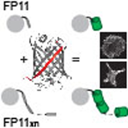
          Tagging proteins with fluorescent proteins is a powerful method for both imaging and non-imaging applications. Here the authors use the eleventh β-strand of sfGFP and sfCherry as epitope tags for multicolour imaging and amplified signals by tandem arrangement; shortness of the tag enabled introduction into genomic loci using CRISPR/Cas9.

          Related collections

          Most cited references19

          • Record: found
          • Abstract: found
          • Article: not found

          Protein tagging and detection with engineered self-assembling fragments of green fluorescent protein.

          Existing protein tagging and detection methods are powerful but have drawbacks. Split protein tags can perturb protein solubility or may not work in living cells. Green fluorescent protein (GFP) fusions can misfold or exhibit altered processing. Fluorogenic biarsenical FLaSH or ReASH substrates overcome many of these limitations but require a polycysteine tag motif, a reducing environment and cell transfection or permeabilization. An ideal protein tag would be genetically encoded, would work both in vivo and in vitro, would provide a sensitive analytical signal and would not require external chemical reagents or substrates. One way to accomplish this might be with a split GFP, but the GFP fragments reported thus far are large and fold poorly, require chemical ligation or fused interacting partners to force their association, or require coexpression or co-refolding to produce detectable folded and fluorescent GFP. We have engineered soluble, self-associating fragments of GFP that can be used to tag and detect either soluble or insoluble proteins in living cells or cell lysates. The split GFP system is simple and does not change fusion protein solubility.
            Bookmark
            • Record: found
            • Abstract: found
            • Article: not found

            Fluorescent probes for super-resolution imaging in living cells.

            In 1873, Ernst Abbe discovered that features closer than approximately 200 nm cannot be resolved by lens-based light microscopy. In recent years, however, several new far-field super-resolution imaging techniques have broken this diffraction limit, producing, for example, video-rate movies of synaptic vesicles in living neurons with 62 nm spatial resolution. Current research is focused on further improving spatial resolution in an effort to reach the goal of video-rate imaging of live cells with molecular (1-5 nm) resolution. Here, we describe the contributions of fluorescent probes to far-field super-resolution imaging, focusing on fluorescent proteins and organic small-molecule fluorophores. We describe the features of existing super-resolution fluorophores and highlight areas of importance for future research and development.
              Bookmark
              • Record: found
              • Abstract: found
              • Article: not found

              GFP Reconstitution Across Synaptic Partners (GRASP) defines cell contacts and synapses in living nervous systems.

              The identification of synaptic partners is challenging in dense nerve bundles, where many processes occupy regions beneath the resolution of conventional light microscopy. To address this difficulty, we have developed GRASP, a system to label membrane contacts and synapses between two cells in living animals. Two complementary fragments of GFP are expressed on different cells, tethered to extracellular domains of transmembrane carrier proteins. When the complementary GFP fragments are fused to ubiquitous transmembrane proteins, GFP fluorescence appears uniformly along membrane contacts between the two cells. When one or both GFP fragments are fused to synaptic transmembrane proteins, GFP fluorescence is tightly localized to synapses. GRASP marks known synaptic contacts in C. elegans, correctly identifies changes in mutants with altered synaptic specificity, and can uncover new information about synaptic locations as confirmed by electron microscopy. GRASP may prove particularly useful for defining connectivity in complex nervous systems.
                Bookmark

                Author and article information

                Journal
                Nat Commun
                Nat Commun
                Nature Communications
                Nature Publishing Group
                2041-1723
                18 March 2016
                2016
                : 7
                : 11046
                Affiliations
                [1 ]Department of Pharmaceutical Chemistry, University of California , San Francisco, California 94143, USA
                [2 ]Tetrad Graduate Program, University of California , San Francisco, California 94143, USA
                [3 ]Department of Cellular and Molecular Pharmacology, University of California , San Francisco, California 94143, USA
                [4 ]Department of Biochemistry and Biophysics, University of California , San Francisco, California 94143, USA
                [5 ]Howard Hughes Medical Institute , San Francisco, California 94143, USA
                Author notes
                [*]

                These authors contributed equally to this work

                Article
                ncomms11046
                10.1038/ncomms11046
                4802074
                26988139
                41ac1208-ab9f-49ec-a1ec-10c915e52801
                Copyright © 2016, Nature Publishing Group, a division of Macmillan Publishers Limited. All Rights Reserved.

                This work is licensed under a Creative Commons Attribution 4.0 International License. The images or other third party material in this article are included in the article's Creative Commons license, unless indicated otherwise in the credit line; if the material is not included under the Creative Commons license, users will need to obtain permission from the license holder to reproduce the material. To view a copy of this license, visit http://creativecommons.org/licenses/by/4.0/

                History
                : 11 December 2015
                : 15 February 2016
                Categories
                Article

                Uncategorized
                Uncategorized

                Comments

                Comment on this article