13
views
0
recommends
+1 Recommend
0 collections
    0
    shares
      • Record: found
      • Abstract: found
      • Article: found
      Is Open Access

      Improved split fluorescent proteins for endogenous protein labeling

      research-article

      Read this article at

      Bookmark
          There is no author summary for this article yet. Authors can add summaries to their articles on ScienceOpen to make them more accessible to a non-specialist audience.

          Abstract

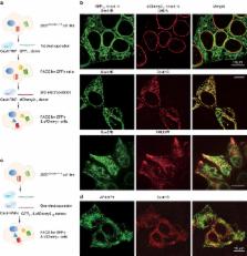
          Self-complementing split fluorescent proteins (FPs) have been widely used for protein labeling, visualization of subcellular protein localization, and detection of cell–cell contact. To expand this toolset, we have developed a screening strategy for the direct engineering of self-complementing split FPs. Via this strategy, we have generated a yellow–green split-mNeonGreen2 1–10/11 that improves the ratio of complemented signal to the background of FP 1–10-expressing cells compared to the commonly used split GFP 1–10/11; as well as a 10-fold brighter red-colored split-sfCherry2 1–10/11. Based on split sfCherry2, we have engineered a photoactivatable variant that enables single-molecule localization-based super-resolution microscopy. We have demonstrated dual-color endogenous protein tagging with sfCherry2 11 and GFP 11, revealing that endoplasmic reticulum translocon complex Sec61B has reduced abundance in certain peripheral tubules. These new split FPs not only offer multiple colors for imaging interaction networks of endogenous proteins, but also hold the potential to provide orthogonal handles for biochemical isolation of native protein complexes.

          Abstract

          Split fluorescent proteins (FPs) have been widely used to visualise proteins in cells. Here the authors develop a screen for engineering new split FPs, and report a yellow-green split-mNeonGreen2 with reduced background, a red split-sfCherry2 for multicolour labeling, and its photoactivatable variant for super-resolution use.

          Related collections

          Most cited references32

          • Record: found
          • Abstract: found
          • Article: not found

          Three-dimensional super-resolution imaging by stochastic optical reconstruction microscopy.

          Recent advances in far-field fluorescence microscopy have led to substantial improvements in image resolution, achieving a near-molecular resolution of 20 to 30 nanometers in the two lateral dimensions. Three-dimensional (3D) nanoscale-resolution imaging, however, remains a challenge. We demonstrated 3D stochastic optical reconstruction microscopy (STORM) by using optical astigmatism to determine both axial and lateral positions of individual fluorophores with nanometer accuracy. Iterative, stochastic activation of photoswitchable probes enables high-precision 3D localization of each probe, and thus the construction of a 3D image, without scanning the sample. Using this approach, we achieved an image resolution of 20 to 30 nanometers in the lateral dimensions and 50 to 60 nanometers in the axial dimension. This development allowed us to resolve the 3D morphology of nanoscopic cellular structures.
            Bookmark
            • Record: found
            • Abstract: found
            • Article: not found

            A bright monomeric green fluorescent protein derived from Branchiostoma lanceolatum

            Despite the existence of fluorescent proteins spanning the entire visual spectrum, the bulk of modern imaging experiments continue to rely on variants of the green fluorescent protein derived from Aequorea victoria. Meanwhile, a great deal of recent effort has been devoted to engineering and improving red fluorescent proteins, and relatively little attention has been given to green and yellow variants. Here we report a novel monomeric yellow-green fluorescent protein, mNeonGreen, which is derived from a tetrameric fluorescent protein from the cephalochordate Branchiostoma lanceolatum. This fluorescent protein is the brightest monomeric green or yellow fluorescent protein yet described, performs exceptionally well as a fusion tag for traditional imaging as well as stochastic single-molecule superresolution imaging, and is an excellent FRET acceptor for the newest generation of cyan fluorescent proteins.
              Bookmark
              • Record: found
              • Abstract: found
              • Article: not found

              Visualization of interactions among bZIP and Rel family proteins in living cells using bimolecular fluorescence complementation.

              Networks of protein interactions coordinate cellular functions. We describe a bimolecular fluorescence complementation (BiFC) assay for determination of the locations of protein interactions in living cells. This approach is based on complementation between two nonfluorescent fragments of the yellow fluorescent protein (YFP) when they are brought together by interactions between proteins fused to each fragment. BiFC analysis was used to investigate interactions among bZIP and Rel family transcription factors. Regions outside the bZIP domains determined the locations of bZIP protein interactions. The subcellular sites of protein interactions were regulated by signaling. Cross-family interactions between bZIP and Rel proteins affected their subcellular localization and modulated transcription activation. These results attest to the general applicability of the BiFC assay for studies of protein interactions.
                Bookmark

                Author and article information

                Contributors
                bo.huang@ucsf.edu
                Journal
                Nat Commun
                Nat Commun
                Nature Communications
                Nature Publishing Group UK (London )
                2041-1723
                29 August 2017
                29 August 2017
                2017
                : 8
                : 370
                Affiliations
                [1 ]ISNI 0000 0001 2181 7878, GRID grid.47840.3f, , The UC Berkeley-UCSF Graduate Program in Bioengineering, ; San Francisco, CA 94143 USA
                [2 ]ISNI 0000 0001 2297 6811, GRID grid.266102.1, Department of Pharmaceutical Chemistry, , University of California in San Francisco, ; San Francisco, CA 94143 USA
                [3 ]ISNI 0000 0001 2297 6811, GRID grid.266102.1, Graduate Program of Biophysics, , University of California, San Francisco, ; San Francisco, CA 94143 USA
                [4 ]ISNI 0000 0001 2297 6811, GRID grid.266102.1, Department of Cellular and Molecular Pharmacology, and Howard Hughes Medical Institute, , University of California, San Francisco, ; San Francisco, CA 94143 USA
                [5 ]ISNI 0000 0001 2297 6811, GRID grid.266102.1, Department of Biochemistry and Biophysics, , University of California, San Francisco, ; San Francisco, CA 94143 USA
                [6 ]Chan Zuckerberg Biohub, San Francisco, CA 94158 USA
                Author information
                http://orcid.org/0000-0003-1704-4141
                Article
                494
                10.1038/s41467-017-00494-8
                5575300
                28851864
                6038b3fb-d0db-4bd5-b946-7b4d3077c4a8
                © The Author(s) 2017

                Open Access This article is licensed under a Creative Commons Attribution 4.0 International License, which permits use, sharing, adaptation, distribution and reproduction in any medium or format, as long as you give appropriate credit to the original author(s) and the source, provide a link to the Creative Commons license, and indicate if changes were made. The images or other third party material in this article are included in the article’s Creative Commons license, unless indicated otherwise in a credit line to the material. If material is not included in the article’s Creative Commons license and your intended use is not permitted by statutory regulation or exceeds the permitted use, you will need to obtain permission directly from the copyright holder. To view a copy of this license, visit http://creativecommons.org/licenses/by/4.0/.

                History
                : 9 November 2016
                : 30 June 2017
                Categories
                Article
                Custom metadata
                © The Author(s) 2017

                Uncategorized
                Uncategorized

                Comments

                Comment on this article