10
views
0
recommends
+1 Recommend
1 collections
    0
    shares
      • Record: found
      • Abstract: found
      • Article: found
      Is Open Access

      The LIM protein complex establishes a retinal circuitry of visual adaptation by regulating Pax6 α-enhancer activity

      research-article

      Read this article at

      Bookmark
          There is no author summary for this article yet. Authors can add summaries to their articles on ScienceOpen to make them more accessible to a non-specialist audience.

          Abstract

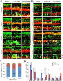
          The visual responses of vertebrates are sensitive to the overall composition of retinal interneurons including amacrine cells, which tune the activity of the retinal circuitry. The expression of Paired-homeobox 6 (PAX6) is regulated by multiple cis-DNA elements including the intronic α-enhancer, which is active in GABAergic amacrine cell subsets. Here, we report that the transforming growth factor ß1-induced transcript 1 protein (Tgfb1i1) interacts with the LIM domain transcription factors Lhx3 and Isl1 to inhibit the α-enhancer in the post-natal mouse retina. Tgfb1i1 -/- mice show elevated α-enhancer activity leading to overproduction of Pax6ΔPD isoform that supports the GABAergic amacrine cell fate maintenance. Consequently, the Tgfb1i1 -/- mouse retinas show a sustained light response, which becomes more transient in mice with the auto-stimulation-defective Pax6 ΔPBS/ΔPBS mutation. Together, we show the antagonistic regulation of the α-enhancer activity by Pax6 and the LIM protein complex is necessary for the establishment of an inner retinal circuitry, which controls visual adaptation.

          DOI: http://dx.doi.org/10.7554/eLife.21303.001

          eLife digest

          The retina is a light-sensitive layer of tissue that lines the inside of the eye. This tissue is highly organized and comprises a variety of different nerve cells, including amacrine cells. Together, these cells process incoming light and then trigger electrical signals that travel to the brain, where they are translated into an image. Changes in the nerve cell composition of the retina, or in how the cells connect to each other, can alter the visual information that travels to the brain.

          The nerve cells of the retina are formed before a young animal opens its eyes for the first time. Proteins called transcription factors – which regulate the expression of genes – tightly control how the retina develops. For example, a transcription factor called Pax6 drives the development of amacrine cells. Several other transcription factors control the production of Pax6 by binding to a section of DNA known as the “α-enhancer”. However, it is not clear how regulating Pax6 production influences the development of specific sets of amacrine cells.

          Kim et al. reveal that a protein known as Tgfb1i1 interacts with two transcription factors to form a “complex” that binds to the α-enhancer and blocks the production of a particular form of Pax6. In experiments performed in mice, the loss of Tgfb1i1 led to increased production of this form of Pax6, which resulted in the retina containing more of a certain type of amacrine cell that produce a molecule called GABA. Mice lacking Tgfb1i1 show a stronger response to light and are therefore comparable to people who are too sensitive to light. On the other hand, mice with a missing a section of the α-enhancer DNA have fewer amacrine cells releasing GABA and become less sensitive to light and are comparable to people who have difficulty detecting weaker light signals.

          The findings of Kim et al. suggest that an individual’s sensitivity to light is related, at least in part, to the mixture of amacrine cells found in their retina, which is determined by certain transcription factors that target the α-enhancer.

          DOI: http://dx.doi.org/10.7554/eLife.21303.002

          Related collections

          Most cited references52

          • Record: found
          • Abstract: found
          • Article: not found

          Electroporation and RNA interference in the rodent retina in vivo and in vitro.

          The large number of candidate genes made available by comprehensive genome analysis requires that relatively rapid techniques for the study of function be developed. Here, we report a rapid and convenient electroporation method for both gain- and loss-of-function studies in vivo and in vitro in the rodent retina. Plasmid DNA directly injected into the subretinal space of neonatal rodent pups was taken up by a significant fraction of exposed cells after several pulses of high voltage. With this technique, GFP expression vectors were efficiently transfected into retinal cells with little damage to the operated pups. Transfected GFP allowed clear visualization of cell morphologies, and the expression persisted for at least 50 days. DNA-based RNA interference vectors directed against two transcription factors important in photoreceptor development led to photoreceptor phenotypes similar to those of the corresponding knockout mice. Reporter constructs carrying retinal cell type-specific promoters were readily introduced into the retina in vivo, where they exhibited the appropriate expression patterns. Plasmid DNA was also efficiently transfected into retinal explants in vitro by high-voltage pulses.
            Bookmark
            • Record: found
            • Abstract: found
            • Article: not found

            Pax6 is required for the multipotent state of retinal progenitor cells.

            The molecular mechanisms mediating the retinogenic potential of multipotent retinal progenitor cells (RPCs) are poorly defined. Prior to initiating retinogenesis, RPCs express a limited set of transcription factors implicated in the evolutionary ancient genetic network that initiates eye development. We elucidated the function of one of these factors, Pax6, in the RPCs of the intact developing eye by conditional gene targeting. Upon Pax6 inactivation, the potential of RPCs becomes entirely restricted to only one of the cell fates normally available to RPCs, resulting in the exclusive generation of amacrine interneurons. Our findings demonstrate furthermore that Pax6 directly controls the transcriptional activation of retinogenic bHLH factors that bias subsets of RPCs toward the different retinal cell fates, thereby mediating the full retinogenic potential of RPCs.
              Bookmark
              • Record: found
              • Abstract: found
              • Article: not found

              Otx2 homeobox gene controls retinal photoreceptor cell fate and pineal gland development.

              Understanding the molecular mechanisms by which distinct cell fate is determined during organogenesis is a central issue in development and disease. Here, using conditional gene ablation in mice, we show that the transcription factor Otx2 is essential for retinal photoreceptor cell fate determination and development of the pineal gland. Otx2-deficiency converted differentiating photoreceptor cells to amacrine-like neurons and led to a total lack of pinealocytes in the pineal gland. We also found that Otx2 transactivates the cone-rod homeobox gene Crx, which is required for terminal differentiation and maintenance of photoreceptor cells. Furthermore, retroviral gene transfer of Otx2 steers retinal progenitor cells toward becoming photoreceptors. Thus, Otx2 is a key regulatory gene for the cell fate determination of retinal photoreceptor cells. Our results reveal the key molecular steps required for photoreceptor cell-fate determination and pinealocyte development.
                Bookmark

                Author and article information

                Contributors
                Role: Reviewing editor
                Journal
                eLife
                Elife
                eLife
                eLife
                eLife
                eLife Sciences Publications, Ltd
                2050-084X
                31 January 2017
                2017
                : 6
                : e21303
                Affiliations
                [1 ]deptDepartment of Biological Sciences , Korea Advanced Institute of Science and Technology (KAIST) , Daejeon, South Korea
                [2 ]deptDepartment of Physiology , Chungbuk National University School of Medicine , Cheongju, South Korea
                [3 ]deptDepartment of Anatomy, College of Medicine , The Catholic University of Korea , Seoul, South Korea
                [4 ]deptDepartment of Biochemistry , Showa University School of Medicine , Tokyo, Japan
                [5 ]deptDepartment of Life Sciences , Gwangju Institute of Science and Technology (GIST) , Gwangju, South Korea
                [6 ]deptDepartment of Ophthalmology and Visual Sciences , Vanderbilt University , Nashville, United States
                [7 ]KAIST Institute of BioCentury , Daejeon, South Korea
                [8]Johns Hopkins University School of Medicine , United States
                [9]Johns Hopkins University School of Medicine , United States
                Author notes
                [†]

                These authors contributed equally to this work.

                Author information
                http://orcid.org/0000-0002-9486-5771
                http://orcid.org/0000-0003-0767-1918
                Article
                21303
                10.7554/eLife.21303
                5308899
                28139974
                44f9fb30-a773-4970-81db-6261d43cde11
                © 2017, Kim et al

                This article is distributed under the terms of the Creative Commons Attribution License, which permits unrestricted use and redistribution provided that the original author and source are credited.

                History
                : 06 September 2016
                : 23 January 2017
                Funding
                Funded by: FundRef http://dx.doi.org/10.13039/501100003725, National Research Foundation of Korea;
                Award ID: NRF-2009-00424
                Award Recipient :
                Funded by: FundRef http://dx.doi.org/10.13039/501100001321, National Research Foundation;
                Award ID: NRF-2006-2004289
                Award Recipient :
                Funded by: FundRef http://dx.doi.org/10.13039/100000053, National Eye Institute;
                Award ID: NIH R01-EY013760
                Award Recipient :
                Funded by: FundRef http://dx.doi.org/10.13039/501100003725, National Research Foundation of Korea;
                Award ID: NRF-2013-056566
                Award Recipient :
                Funded by: FundRef http://dx.doi.org/10.13039/501100003725, National Research Foundation of Korea;
                Award ID: NRF-2014R1A2A2A01003069
                Award Recipient :
                The funders had no role in study design, data collection and interpretation, or the decision to submit the work for publication.
                Categories
                Research Article
                Genes and Chromosomes
                Neuroscience
                Custom metadata
                2.5
                Regulation of Pax6 expression through the α-enhancer fine-tunes amacrine cell subtype number, and consequently, the visual output of the retina.

                Life sciences
                pax6,lim domain protein,retina,cell fate determination,visual adaptation,mouse
                Life sciences
                pax6, lim domain protein, retina, cell fate determination, visual adaptation, mouse

                Comments

                Comment on this article