Inviting an author to review:
Find an author and click ‘Invite to review selected article’ near their name.
Search for authorsSearch for similar articles
23
views
0
recommends
+1 Recommend
0 collections
    0
    shares
      • Record: found
      • Abstract: found
      • Article: found
      Is Open Access

      Cryo-EM structure of Mcm2-7 double hexamer on DNA suggests a lagging-strand DNA extrusion model

      research-article

      Read this article at

      Bookmark
          There is no author summary for this article yet. Authors can add summaries to their articles on ScienceOpen to make them more accessible to a non-specialist audience.

          Significance

          During initiation of DNA replication in eukaryotes, the origin recognition complex, with Cdc6 and Cdt1, assembles an inactive Mcm2-7 double hexamer on the dsDNA. Later, the double hexamer recruits Cdc45 and GINS to form two active and separate DNA helicases. The active Cdc45–Mcm2-7–GINS helicase encircles the leading strand while excluding the lagging strand. One of the fundamental unanswered questions is how each Mcm2-7 hexamer converts from binding dsDNA to binding one of the single strands. The structure of the double hexamer on dsDNA reveals how DNA interacts with key elements inside the central channel, leading us to propose a lagging-strand extrusion mechanism. This work advances our understanding of eukaryotic replication initiation.

          Abstract

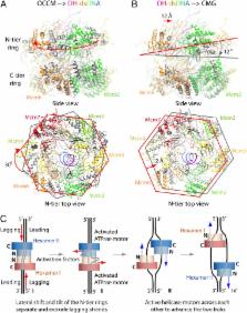
          During replication initiation, the core component of the helicase—the Mcm2-7 hexamer—is loaded on origin DNA as a double hexamer (DH). The two ring-shaped hexamers are staggered, leading to a kinked axial channel. How the origin DNA interacts with the axial channel is not understood, but the interaction could provide key insights into Mcm2-7 function and regulation. Here, we report the cryo-EM structure of the Mcm2-7 DH on dsDNA and show that the DNA is zigzagged inside the central channel. Several of the Mcm subunit DNA-binding loops, such as the oligosaccharide–oligonucleotide loops, helix 2 insertion loops, and presensor 1 (PS1) loops, are well defined, and many of them interact extensively with the DNA. The PS1 loops of Mcm 3, 4, 6, and 7, but not 2 and 5, engage the lagging strand with an approximate step size of one base per subunit. Staggered coupling of the two opposing hexamers positions the DNA right in front of the two Mcm2–Mcm5 gates, with each strand being pressed against one gate. The architecture suggests that lagging-strand extrusion initiates in the middle of the DH that is composed of the zinc finger domains of both hexamers. To convert the Mcm2-7 DH structure into the Mcm2-7 hexamer structure found in the active helicase, the N-tier ring of the Mcm2-7 hexamer in the DH-dsDNA needs to tilt and shift laterally. We suggest that these N-tier ring movements cause the DNA strand separation and lagging-strand extrusion.

          Related collections

          Most cited references86

          • Record: found
          • Abstract: found
          • Article: not found

          DNA replication in eukaryotic cells.

          The maintenance of the eukaryotic genome requires precisely coordinated replication of the entire genome each time a cell divides. To achieve this coordination, eukaryotic cells use an ordered series of steps to form several key protein assemblies at origins of replication. Recent studies have identified many of the protein components of these complexes and the time during the cell cycle they assemble at the origin. Interestingly, despite distinct differences in origin structure, the identity and order of assembly of eukaryotic replication factors is highly conserved across all species. This review describes our current understanding of these events and how they are coordinated with cell cycle progression. We focus on bringing together the results from different organisms to provide a coherent model of the events of initiation. We emphasize recent progress in determining the function of the different replication factors once they have been assembled at the origin.
            Bookmark
            • Record: found
            • Abstract: found
            • Article: not found

            Concerted loading of Mcm2-7 double hexamers around DNA during DNA replication origin licensing.

            The licensing of eukaryotic DNA replication origins, which ensures once-per-cell-cycle replication, involves the loading of six related minichromosome maintenance proteins (Mcm2-7) into prereplicative complexes (pre-RCs). Mcm2-7 forms the core of the replicative DNA helicase, which is inactive in the pre-RC. The loading of Mcm2-7 onto DNA requires the origin recognition complex (ORC), Cdc6, and Cdt1, and depends on ATP. We have reconstituted Mcm2-7 loading with purified budding yeast proteins. Using biochemical approaches and electron microscopy, we show that single heptamers of Cdt1*Mcm2-7 are loaded cooperatively and result in association of stable, head-to-head Mcm2-7 double hexamers connected via their N-terminal rings. DNA runs through a central channel in the double hexamer, and, once loaded, Mcm2-7 can slide passively along double-stranded DNA. Our work has significant implications for understanding how eukaryotic DNA replication origins are chosen and licensed, how replisomes assemble during initiation, and how unwinding occurs during DNA replication.
              Bookmark
              • Record: found
              • Abstract: found
              • Article: not found

              Regulated Eukaryotic DNA Replication Origin Firing with Purified Proteins

              Eukaryotic cells initiate DNA replication from multiple origins, which must be tightly regulated to promote precise genome duplication in every cell cycle. To accomplish this, initiation is partitioned into two temporally discrete steps: a double hexameric MCM complex is first loaded at replication origins during G1 phase, and then converted to the active CMG (Cdc45, MCM, GINS) helicase during S phase. Here we describe the reconstitution of budding yeast DNA replication initiation with 16 purified replication factors, made from 42 polypeptides. Origin-dependent initiation recapitulates regulation seen in vivo. Cyclin dependent kinase (CDK) inhibits MCM loading by phosphorylating the origin recognition complex (ORC) and promotes CMG formation by phosphorylating Sld2 and Sld3. Dbf4 dependent kinase (DDK) promotes replication by phosphorylating MCM, and can act either before or after CDK. These experiments define the minimum complement of proteins, protein kinase substrates and co-factors required for regulated eukaryotic DNA replication.
                Bookmark

                Author and article information

                Journal
                Proc Natl Acad Sci U S A
                Proc. Natl. Acad. Sci. U.S.A
                pnas
                pnas
                PNAS
                Proceedings of the National Academy of Sciences of the United States of America
                National Academy of Sciences
                0027-8424
                1091-6490
                7 November 2017
                25 October 2017
                25 October 2017
                : 114
                : 45
                : E9529-E9538
                Affiliations
                [1] aDNA Replication Group, Institute of Clinical Sciences, Faculty of Medicine, Imperial College London , London W12 0NN, United Kingdom;
                [2] bMedical Research Council London Institute of Medical Sciences , London W12 0NN, United Kingdom;
                [3] cCryo-EM Structural Biology Laboratory, Van Andel Research Institute , Grand Rapids, MI 49503;
                [4] dCold Spring Harbor Laboratory , Cold Spring Harbor, NY11724
                Author notes
                2To whom correspondence may be addressed. Email: stillman@ 123456cshl.edu , chris.speck@ 123456imperial.ac.uk , or Huilin.Li@ 123456VAI.org .

                Contributed by Bruce Stillman, September 20, 2017 (sent for review July 16, 2017; reviewed by Stephen Bell and Eric J. Enemark)

                Author contributions: Y.N., B.S., C.S., and H.L. designed research; Y.N., Z.Y., L.B., S.S., and G.Z. performed research; Y.N., Z.Y., L.B., S.S., G.Z., B.S., C.S., and H.L. analyzed data; and B.S., C.S., and H.L. wrote the paper.

                Reviewers: S.B., Howard Hughes Medical Institute, MIT; and E.J.E., St. Jude Children’s Research Hospital.

                1Y.N., Z.Y., and L.B. contributed equally to this work.

                Author information
                http://orcid.org/0000-0002-9453-4091
                http://orcid.org/0000-0001-6646-1692
                Article
                201712537
                10.1073/pnas.1712537114
                5692578
                29078375
                45c9711c-281a-490c-bbd7-2aeca1a47ce9
                Copyright © 2017 the Author(s). Published by PNAS.

                This open access article is distributed under Creative Commons Attribution-NonCommercial-NoDerivatives License 4.0 (CC BY-NC-ND).

                History
                Page count
                Pages: 10
                Funding
                Funded by: HHS | NIH | National Institute of General Medical Sciences (NIGMS) 100000057
                Award ID: GM111472
                Funded by: HHS | NIH | National Institute of General Medical Sciences (NIGMS) 100000057
                Award ID: GM45436
                Funded by: RCUK | Biotechnology and Biological Sciences Research Council (BBSRC) 501100000268
                Award ID: BB/N000323/1
                Funded by: Wellcome Trust
                Award ID: 107903/Z/15/Z
                Funded by: RCUK | Medical Research Council (MRC) 501100000265
                Award ID: MC_U120085811
                Funded by: Van Andel Research Institute (VARI) 100006019
                Award ID: None
                Categories
                PNAS Plus
                Biological Sciences
                Biochemistry
                PNAS Plus

                dna replication,helicase,dna unwinding,mini chromosome maintenance,cryo-electron microscopy

                Comments

                Comment on this article