11
views
0
recommends
+1 Recommend
0 collections
    0
    shares
      • Record: found
      • Abstract: found
      • Article: found
      Is Open Access

      Colour Vignetting Correction for Microscopy Image Mosaics Used for Quantitative Analyses

      research-article
      1 , 2 , 3 ,
      BioMed Research International
      Hindawi

      Read this article at

      Bookmark
          There is no author summary for this article yet. Authors can add summaries to their articles on ScienceOpen to make them more accessible to a non-specialist audience.

          Abstract

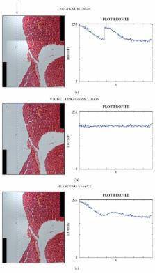
          Image mosaicing permits achieving one high-resolution image, extending the visible area of the sample while keeping the same resolution. However, intensity inhomogeneity of the stitched images can alter measurements and the right perception of the original sample. The problem can be solved by flat-field correcting the images through the vignetting function. Vignetting correction has been widely addressed for grey-level images, but not for colour ones. In this work, a practical solution for the colour vignetting correction in microscopy, also facing the problem of saturated pixels, is described. In order to assess the quality of the proposed approach, five different tonal correction approaches were quantitatively compared using state-of-the-art metrics and seven pairs of partially overlapping images of seven different samples. The results obtained proved that the proposed approach allows obtaining high quality colour flat-field corrected images and seamless mosaics without employing any blending adjustment. In order to give the opportunity to easily obtain seamless mosaics ready for quantitative analysis, the described vignetting correction method has been implemented in an upgraded release of MicroMos (version 3.0), an open-source software specifically designed to automatically obtain mosaics of partially overlapped images.

          Related collections

          Most cited references57

          • Record: found
          • Abstract: not found
          • Article: not found

          A universal image quality index

            Bookmark
            • Record: found
            • Abstract: not found
            • Article: not found

            Automatic Panoramic Image Stitching using Invariant Features

              Bookmark
              • Record: found
              • Abstract: found
              • Article: not found

              MTT assay for cell viability: Intracellular localization of the formazan product is in lipid droplets.

              Although MTT is widely used to assess cytotoxicity and cell viability, the precise localization of its reduced formazan product is still unclear. In the present study the localization of MTT formazan was studied by direct microscopic observation of living HeLa cells and by colocalization analysis with organelle-selective fluorescent probes. MTT formazan granules did not colocalize with mitochondria as revealed by rhodamine 123 labeling or autofluorescence. Likewise, no colocalization was observed between MTT formazan granules and lysosomes labeled by neutral red. Taking into account the lipophilic character and lipid solubility of MTT formazan, an evaluation of the MTT reaction was performed after treatment of cells with sunflower oil emulsions to induce a massive occurrence of lipid droplets. Under this condition, lipid droplets revealed a large amount of MTT formazan deposits. Kinetic studies on the viability of MTT-treated cells showed no harmful effects at short times. Quantitative structure-activity relations (QSAR) models were used to predict and explain the localization of both the MTT tetrazolium salt and its formazan product. These predictions were in agreement with experimental observations on the accumulation of MTT formazan product in lipid droplets. Copyright © 2012 Elsevier GmbH. All rights reserved.
                Bookmark

                Author and article information

                Contributors
                Journal
                Biomed Res Int
                Biomed Res Int
                BMRI
                BioMed Research International
                Hindawi
                2314-6133
                2314-6141
                2018
                7 June 2018
                : 2018
                : 7082154
                Affiliations
                1Istituto Scientifico Romagnolo per lo Studio e la Cura dei Tumori (IRST) IRCCS, Meldola, Italy
                2Advanced Research Center on Electronic Systems for Information and Communication Technologies “E. De Castro” (ARCES), University of Bologna, Bologna, Italy
                3Department of Computer Science and Engineering (DISI), University of Bologna, Bologna, Italy
                Author notes

                Academic Editor: Jiang Du

                Author information
                http://orcid.org/0000-0003-2938-5058
                Article
                10.1155/2018/7082154
                6011154
                4725faaf-5786-4a98-9824-3ee0564d72b7
                Copyright © 2018 Filippo Piccinini and Alessandro Bevilacqua.

                This is an open access article distributed under the Creative Commons Attribution License, which permits unrestricted use, distribution, and reproduction in any medium, provided the original work is properly cited.

                History
                : 21 February 2018
                : 30 April 2018
                : 10 May 2018
                Funding
                Funded by: IRST IRCCS
                Funded by: Università di Bologna
                Categories
                Research Article

                Comments

                Comment on this article