39
views
0
recommends
+1 Recommend
0 collections
    0
    shares
      • Record: found
      • Abstract: found
      • Article: found
      Is Open Access

      The Nucleocapsid Protein of Rift Valley Fever Virus Is a Potent Human CD8 + T Cell Antigen and Elicits Memory Responses

      research-article

      Read this article at

      Bookmark
          There is no author summary for this article yet. Authors can add summaries to their articles on ScienceOpen to make them more accessible to a non-specialist audience.

          Abstract

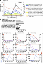
          There is no licensed human vaccine currently available for Rift Valley Fever Virus (RVFV), a Category A high priority pathogen and a serious zoonotic threat. While neutralizing antibodies targeting the viral glycoproteins are protective, they appear late in the course of infection, and may not be induced in time to prevent a natural or bioterrorism-induced outbreak. Here we examined the immunogenicity of RVFV nucleocapsid (N) protein as a CD8 + T cell antigen with the potential for inducing rapid protection after vaccination. HLA-A*0201 (A2)-restricted epitopic determinants were identified with N-specific CD8 + T cells from eight healthy donors that were primed with dendritic cells transduced to express N, and subsequently expanded in vitro by weekly re-stimulations with monocytes pulsed with 59 15mer overlapping peptides (OLPs) across N. Two immunodominant epitopes, VT9 (VLSEWLPVT, N 121–129) and IL9 (ILDAHSLYL, N 165–173), were defined. VT9- and IL9-specific CD8 + T cells identified by tetramer staining were cytotoxic and polyfunctional, characteristics deemed important for viral control in vivo. These peptides induced specific CD8 + T cell responses in A2-transgenic mice, and more importantly, potent N-specific CD8 + T cell reactivities, including VT9- and IL9-specific ones, were mounted by mice after a booster vaccination with the live attenuated RVF MP-12. Our data suggest that the RVFV N protein is a potent human T cell immunogen capable of eliciting broad, immunodominant CD8 + T cell responses that are potentially protective. Understanding the immune responses to the nucleocapsid is central to the design of an effective RVFV vaccine irrespective of whether this viral protein is effective as a stand-alone immunogen or only in combination with other RVFV antigens.

          Related collections

          Most cited references68

          • Record: found
          • Abstract: found
          • Article: not found

          HIV nonprogressors preferentially maintain highly functional HIV-specific CD8+ T cells.

          Establishing a CD8(+) T cell-mediated immune correlate of protection in HIV disease is crucial to the development of vaccines designed to generate cell-mediated immunity. Historically, neither the quantity nor breadth of the HIV-specific CD8(+) T-cell response has correlated conclusively with protection. Here, we assess the quality of the HIV-specific CD8(+) T-cell response by measuring 5 CD8(+) T-cell functions (degranulation, IFN-gamma, MIP-1beta, TNF-alpha, and IL-2) simultaneously in chronically HIV-infected individuals and elite nonprogressors. We find that the functional profile of HIV-specific CD8(+) T cells in progressors is limited compared to that of nonprogressors, who consistently maintain highly functional CD8(+) T cells. This limited functionality is independent of HLA type and T-cell memory phenotype, is HIV-specific rather than generalized, and is not effectively restored by therapeutic intervention. Whereas the total HIV-specific CD8(+) T-cell frequency did not correlate with viral load, the frequency and proportion of the HIV-specific T-cell response with highest functionality inversely correlated with viral load in the progressors. Thus, rather than quantity or phenotype, the quality of the CD8(+) T-cell functional response serves as an immune correlate of HIV disease progression and a potential qualifying factor for evaluation of HIV vaccine efficacy.
            Bookmark
            • Record: found
            • Abstract: found
            • Article: not found

            Plasmacytoid dendritic cells in immunity.

            Human and mouse plasmacytoid dendritic cells have been shown to correspond to a specialized cell population that produces large amounts of type I interferons in response to viruses, the so-called natural interferon-producing cells. As a result, intensive investigation is now focused on the potential functions of plasmacytoid dendritic cells in both innate and adaptive immunity. Here we review recent progress on the characterization of plasmacytoid dendritic cell origin, development, migration and function in immunity and tolerance, as well as their effect on human diseases.
              Bookmark
              • Record: found
              • Abstract: found
              • Article: found
              Is Open Access

              The first T cell response to transmitted/founder virus contributes to the control of acute viremia in HIV-1 infection

              Identification of the transmitted/founder virus makes possible, for the first time, a genome-wide analysis of host immune responses against the infecting HIV-1 proteome. A complete dissection was made of the primary HIV-1–specific T cell response induced in three acutely infected patients. Cellular assays, together with new algorithms which identify sites of positive selection in the virus genome, showed that primary HIV-1–specific T cells rapidly select escape mutations concurrent with falling virus load in acute infection. Kinetic analysis and mathematical modeling of virus immune escape showed that the contribution of CD8 T cell–mediated killing of productively infected cells was earlier and much greater than previously recognized and that it contributed to the initial decline of plasma virus in acute infection. After virus escape, these first T cell responses often rapidly waned, leaving or being succeeded by T cell responses to epitopes which escaped more slowly or were invariant. These latter responses are likely to be important in maintaining the already established virus set point. In addition to mutations selected by T cells, there were other selected regions that accrued mutations more gradually but were not associated with a T cell response. These included clusters of mutations in envelope that were targeted by NAbs, a few isolated sites that reverted to the consensus sequence, and bystander mutations in linkage with T cell–driven escape.
                Bookmark

                Author and article information

                Contributors
                Role: Editor
                Journal
                PLoS One
                PLoS ONE
                plos
                plosone
                PLoS ONE
                Public Library of Science (San Francisco, USA )
                1932-6203
                2013
                18 March 2013
                : 8
                : 3
                : e59210
                Affiliations
                [1 ]Department of Biological Science and Border Biomedical Research Center, The University of Texas at El Paso, El Paso, Texas, United States of America
                [2 ]Center for Infectious Disease, La Jolla Institute for Allergy and Immunology, La Jolla, California, United States of America
                [3 ]Institute of Infection and Immunity, Cardiff University School of Medicine, Heath Park, Cardiff, United Kingdom
                [4 ]Department of Microbiology and Immunology, The University of Texas Medical Branch, Galveston, Texas, United States of America
                George Mason University, United States of America
                Author notes

                Competing Interests: Co-author Dr. Douglas M. Watts is a PLOS ONE Editorial Board member. This does not alter the authors' adherence to all the PLOS ONE policies on sharing data and materials. The authors have also reviewed the authors’ declaration of potential competing interests for the re-submission according to PLOS ONE policy. All authors declared no competing interests.

                Conceived and designed the experiments: JK-M WX DMW MCC XT. Performed the experiments: WX MCC DMW LAV XT FJ JS. Analyzed the data: WX MCC DMW JCM XT JK-M. Contributed reagents/materials/analysis tools: JS AS SM JCM AKS LW CJP. Wrote the paper: WX MCC JK-M.

                Article
                PONE-D-12-35063
                10.1371/journal.pone.0059210
                3601065
                23527138
                4a2077c9-a567-4b74-bff9-0f1adac463b1
                Copyright @ 2013

                This is an open-access article distributed under the terms of the Creative Commons Attribution License, which permits unrestricted use, distribution, and reproduction in any medium, provided the original author and source are credited.

                History
                : 9 November 2012
                : 12 February 2013
                Page count
                Pages: 12
                Funding
                This work was supported by National Institutes of Health (NIH) research grants AI064069 and AI77413 to June Kan-Mitchell, and U54 AI057156 to David H. Walker. The funders had no role in study design, data collection and analysis, decision to publish, or preparation of the manuscript.
                Categories
                Research Article
                Biology
                Immunology
                Immune Cells
                T Cells
                Microbiology
                Virology
                Viral Structure
                Nucleocapsid
                Viral Vaccines
                Microbial Control
                Model Organisms
                Animal Models
                Mouse
                Medicine
                Clinical Immunology
                Immunity
                Vaccination
                Vaccines
                Vaccine Development
                Infectious Diseases
                Neglected Tropical Diseases
                Rift Valley fever
                Viral Diseases
                Rift Valley fever
                Veterinary Science
                Veterinary Diseases
                Zoonotic Diseases

                Uncategorized
                Uncategorized

                Comments

                Comment on this article