0
views
0
recommends
+1 Recommend
0 collections
    0
    shares
      • Record: found
      • Abstract: found
      • Article: found
      Is Open Access

      Investigating silent pauses in connected speech: integrating linguistic, neuropsychological, and neuroanatomical perspectives across narrative tasks in post-stroke aphasia

      research-article

      Read this article at

      Bookmark
          There is no author summary for this article yet. Authors can add summaries to their articles on ScienceOpen to make them more accessible to a non-specialist audience.

          Abstract

          Introduction

          Silent pauses are regarded as integral components of the temporal organization of speech. However, it has also been hypothesized that they serve as markers for internal cognitive processes, including word access, monitoring, planning, and memory functions. Although existing evidence across various pathological populations underscores the importance of investigating silent pauses’ characteristics, particularly in terms of frequency and duration, there is a scarcity of data within the domain of post-stroke aphasia.

          Methods

          The primary objective of the present study is to scrutinize the frequency and duration of silent pauses in two distinct narrative tasks within a cohort of 32 patients with chronic post-stroke aphasia, in comparison with a control group of healthy speakers. Subsequently, we investigate potential correlation patterns between silent pause measures, i.e., frequency and duration, across the two narrative tasks within the patient group, their performance in neuropsychological assessments, and lesion data.

          Results

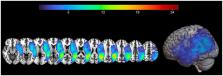
          Our findings showed that patients exhibited a higher frequency of longer-duration pauses in both narrative tasks compared to healthy speakers. Furthermore, within-group comparisons revealed that patients tended to pause more frequently and for longer durations in the picture description task, while healthy participants exhibited the opposite trend. With regard to our second research question, a marginally significant interaction emerged between performance in semantic verbal fluency and the narrative task, in relation to the location of silent pauses—whether between or within clauses—predicting the duration of silent pauses in the patient group. However, no significant results were observed for the frequency of silent pauses. Lastly, our study identified that the duration of silent pauses could be predicted by distinct Regions of Interest (ROIs) in spared tissue within the left hemisphere, as a function of the narrative task.

          Discussion

          Overall, this study follows an integrative approach of linguistic, neuropsychological and neuroanatomical data to define silent pauses in connected speech, and illustrates interrelations between cognitive components, temporal aspects of speech, and anatomical indices, while it further highlights the importance of studying connected speech indices using different narrative tasks.

          Related collections

          Most cited references72

          • Record: found
          • Abstract: not found
          • Article: not found

          Controlling the False Discovery Rate: A Practical and Powerful Approach to Multiple Testing

            Bookmark
            • Record: found
            • Abstract: not found
            • Article: not found

            Fitting Linear Mixed-Effects Models Usinglme4

              Bookmark
              • Record: found
              • Abstract: found
              • Article: not found

              Automated anatomical labeling of activations in SPM using a macroscopic anatomical parcellation of the MNI MRI single-subject brain.

              An anatomical parcellation of the spatially normalized single-subject high-resolution T1 volume provided by the Montreal Neurological Institute (MNI) (D. L. Collins et al., 1998, Trans. Med. Imag. 17, 463-468) was performed. The MNI single-subject main sulci were first delineated and further used as landmarks for the 3D definition of 45 anatomical volumes of interest (AVOI) in each hemisphere. This procedure was performed using a dedicated software which allowed a 3D following of the sulci course on the edited brain. Regions of interest were then drawn manually with the same software every 2 mm on the axial slices of the high-resolution MNI single subject. The 90 AVOI were reconstructed and assigned a label. Using this parcellation method, three procedures to perform the automated anatomical labeling of functional studies are proposed: (1) labeling of an extremum defined by a set of coordinates, (2) percentage of voxels belonging to each of the AVOI intersected by a sphere centered by a set of coordinates, and (3) percentage of voxels belonging to each of the AVOI intersected by an activated cluster. An interface with the Statistical Parametric Mapping package (SPM, J. Ashburner and K. J. Friston, 1999, Hum. Brain Mapp. 7, 254-266) is provided as a freeware to researchers of the neuroimaging community. We believe that this tool is an improvement for the macroscopical labeling of activated area compared to labeling assessed using the Talairach atlas brain in which deformations are well known. However, this tool does not alleviate the need for more sophisticated labeling strategies based on anatomical or cytoarchitectonic probabilistic maps.
                Bookmark

                Author and article information

                Contributors
                URI : https://loop.frontiersin.org/people/436115/overviewRole: Role: Role: Role: Role: Role: Role:
                URI : https://loop.frontiersin.org/people/177803/overviewRole: Role: Role: Role: Role: Role: Role:
                URI : https://loop.frontiersin.org/people/155860/overviewRole: Role: Role: Role:
                URI : https://loop.frontiersin.org/people/885481/overviewRole: Role: Role:
                Role: Role:
                URI : https://loop.frontiersin.org/people/1315671/overviewRole: Role: Role: Role:
                URI : https://loop.frontiersin.org/people/204011/overviewRole: Role: Role: Role:
                Role: Role: Role: Role:
                Role: Role: Role:
                Role: Role: Role:
                URI : https://loop.frontiersin.org/people/915162/overviewRole: Role: Role:
                URI : https://loop.frontiersin.org/people/807367/overviewRole: Role: Role: Role: Role: Role:
                URI : https://loop.frontiersin.org/people/99804/overviewRole: Role: Role: Role:
                URI : https://loop.frontiersin.org/people/33696/overviewRole: Role: Role:
                URI : https://loop.frontiersin.org/people/661111/overviewRole: Role: Role: Role:
                URI : https://loop.frontiersin.org/people/856572/overviewRole: Role: Role: Role: Role: Role: Role: Role:
                Journal
                Front Neurol
                Front Neurol
                Front. Neurol.
                Frontiers in Neurology
                Frontiers Media S.A.
                1664-2295
                12 April 2024
                2024
                : 15
                : 1347514
                Affiliations
                [1] 1Neuropsychology&Language Disorders Unit, 1st Department of Neurology, Eginition Hospital, Faculty of Medicine, National and Kapodistrian University of Athens , Athens, Greece
                [2] 2Department of Psychology, Panteion University of Social and Political Sciences , Athens, Greece
                [3] 3Center for Brain Recovery, Boston University , Boston, MA, United States
                [4] 42nd Department of Radiology, General University Hospital “Attikon”, Medical School, National and Kapodistrian University of Athens , Athens, Greece
                [5] 5The Aphasia Network Lab, Department of Communication Sciences and Disorders, Northeastern University , Boston, MA, United States
                [6] 6School of Medicine, Democritus University of Thrace , Alexandroupolis, Greece
                [7] 7Department of Industrial Design and Production Engineering, School of Engineering, University of West Attica , Athens, Greece
                [8] 8Stroke Unit, 1st Department of Neurology, Eginition Hospital, National and Kapodistrian University of Athens , Athens, Greece
                [9] 9Department of Linguistics, School of Philosophy, National and Kapodistrian University of Athens , Athens, Greece
                [10] 10Department of Neurology and Clinical Neuroscience, University Hospital Freiburg , Freiburg, Germany
                Author notes

                Edited by: John R. Absher, Prisma Health Neuroscience Associates, United States

                Reviewed by: Nomiki Karpathiou, Athens Alzheimer’s Association, Greece

                Marcelo L. Berthier, University of Malaga, Spain

                Xing Huang, Beijing University of Chinese Medicine Third Affiliated Hospital, China

                Stefano F. Cappa, University Institute of Higher Studies in Pavia, Italy

                *Correspondence: G. Angelopoulou, georginangel@ 123456gmail.com
                Article
                10.3389/fneur.2024.1347514
                11047180
                4a216c60-bc3f-4e63-af7b-ef649297e2af
                Copyright © 2024 Angelopoulou, Kasselimis, Varkanitsa, Tsolakopoulos, Papageorgiou, Velonakis, Meier, Karavassilis, Pantoleon, Laskaris, Kelekis, Tountopoulou, Vassilopoulou, Goutsos, Kiran, Weiller, Rijntjes and Potagas.

                This is an open-access article distributed under the terms of the Creative Commons Attribution License (CC BY). The use, distribution or reproduction in other forums is permitted, provided the original author(s) and the copyright owner(s) are credited and that the original publication in this journal is cited, in accordance with accepted academic practice. No use, distribution or reproduction is permitted which does not comply with these terms.

                History
                : 30 November 2023
                : 07 March 2024
                Page count
                Figures: 6, Tables: 7, Equations: 0, References: 74, Pages: 15, Words: 10442
                Funding
                Funded by: Hellenic Foundation for Research and Innovation, doi 10.13039/501100013209;
                The author(s) declare that financial support was received for the research, authorship, and/or publication of this article. The current research work was supported by the Hellenic Foundation for Research and Innovation (H.F.R.I.) under the “First Call for H.F.R.I. Research Projects to support Faculty members and Researchers and the procurement of high-cost research equipment grant” (project number 4081).
                Categories
                Neurology
                Original Research
                Custom metadata
                Applied Neuroimaging

                Neurology
                post-stroke aphasia,silent pauses,stroke story,picture description,neuropsychological performance,dorsal stream,ventral stream

                Comments

                Comment on this article