22
views
0
recommends
+1 Recommend
0 collections
    0
    shares
      • Record: found
      • Abstract: found
      • Article: not found

      Bioactive Silica Nanoparticles Promote Osteoblast Differentiation through Stimulation of Autophagy and Direct Association with LC3 and p62

      research-article

      Read this article at

      ScienceOpenPublisherPMC
      Bookmark
          There is no author summary for this article yet. Authors can add summaries to their articles on ScienceOpen to make them more accessible to a non-specialist audience.

          Abstract

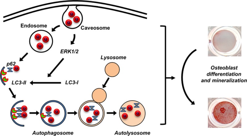
          We recently identified an engineered bioactive silica-based nanoparticle formulation (designated herein as NP1) that stimulates in vitro differentiation and mineralization of osteoblasts, the cells responsible for bone formation, and increases bone mineral density in young mice in vivo. The results demonstrate that these nanoparticles have intrinsic biological activity; however, the intracellular fate and a complete understanding of the mechanism(s) involved remains to be elucidated. Here we investigated the cellular mechanism(s) by which NP1 stimulates differentiation and mineralization of osteoblasts. We show that NP1 enters the cells through a caveolae-mediated endocytosis followed by stimulation of the mitogen activated protein kinase ERK1/2 (p44/p42). Our findings further revealed that NP1 stimulates autophagy including the processing of LC3β-I to LC3β-II, a key protein involved in autophagosome formation, which is dependent on ERK1/2 signaling. Using a variant of NP1 with cobalt ferrite magnetic metal core (NP1-MNP) to pull down associated proteins, we found direct binding of LC3β and p62, two key proteins involved in autophagosome formation, with silica nanoparticles. Interestingly, NP1 specifically interacts with the active and autophagosome associated form of LC3β (LC3β-II). Taken together, the stimulation of autophagy and associated signaling suggests a cellular mechanism for the stimulatory effects of silica nanoparticles on osteoblast differentiation and mineralization.

          Related collections

          Most cited references42

          • Record: found
          • Abstract: found
          • Article: not found

          Guidelines for the use and interpretation of assays for monitoring autophagy.

          In 2008 we published the first set of guidelines for standardizing research in autophagy. Since then, research on this topic has continued to accelerate, and many new scientists have entered the field. Our knowledge base and relevant new technologies have also been expanding. Accordingly, it is important to update these guidelines for monitoring autophagy in different organisms. Various reviews have described the range of assays that have been used for this purpose. Nevertheless, there continues to be confusion regarding acceptable methods to measure autophagy, especially in multicellular eukaryotes. A key point that needs to be emphasized is that there is a difference between measurements that monitor the numbers or volume of autophagic elements (e.g., autophagosomes or autolysosomes) at any stage of the autophagic process vs. those that measure flux through the autophagy pathway (i.e., the complete process); thus, a block in macroautophagy that results in autophagosome accumulation needs to be differentiated from stimuli that result in increased autophagic activity, defined as increased autophagy induction coupled with increased delivery to, and degradation within, lysosomes (in most higher eukaryotes and some protists such as Dictyostelium) or the vacuole (in plants and fungi). In other words, it is especially important that investigators new to the field understand that the appearance of more autophagosomes does not necessarily equate with more autophagy. In fact, in many cases, autophagosomes accumulate because of a block in trafficking to lysosomes without a concomitant change in autophagosome biogenesis, whereas an increase in autolysosomes may reflect a reduction in degradative activity. Here, we present a set of guidelines for the selection and interpretation of methods for use by investigators who aim to examine macroautophagy and related processes, as well as for reviewers who need to provide realistic and reasonable critiques of papers that are focused on these processes. These guidelines are not meant to be a formulaic set of rules, because the appropriate assays depend in part on the question being asked and the system being used. In addition, we emphasize that no individual assay is guaranteed to be the most appropriate one in every situation, and we strongly recommend the use of multiple assays to monitor autophagy. In these guidelines, we consider these various methods of assessing autophagy and what information can, or cannot, be obtained from them. Finally, by discussing the merits and limits of particular autophagy assays, we hope to encourage technical innovation in the field.
            Bookmark
            • Record: found
            • Abstract: found
            • Article: not found

            Mesoporous silica nanoparticles: synthesis, biocompatibility and drug delivery.

            In the past decade, mesoporous silica nanoparticles (MSNs) have attracted more and more attention for their potential biomedical applications. With their tailored mesoporous structure and high surface area, MSNs as drug delivery systems (DDSs) show significant advantages over traditional drug nanocarriers. In this review, we overview the recent progress in the synthesis of MSNs for drug delivery applications. First, we provide an overview of synthesis strategies for fabricating ordered MSNs and hollow/rattle-type MSNs. Then, the in vitro and in vivo biocompatibility and biotranslocation of MSNs are discussed in relation to their chemophysical properties including particle size, surface properties, shape, and structure. The review also highlights the significant achievements in drug delivery using mesoporous silica nanoparticles and their multifunctional counterparts as drug carriers. In particular, the biological barriers for nano-based targeted cancer therapy and MSN-based targeting strategies are discussed. We conclude with our personal perspectives on the directions in which future work in this field might be focused. Copyright © 2012 WILEY-VCH Verlag GmbH & Co. KGaA, Weinheim.
              Bookmark
              • Record: found
              • Abstract: found
              • Article: not found

              Elucidating the mechanism of cellular uptake and removal of protein-coated gold nanoparticles of different sizes and shapes.

              We investigated the mechanism by which transferrin-coated gold nanoparticles (Au NP) of different sizes and shapes entered mammalian cells. We determined that transferrin-coated Au NP entered the cells via clathrin-mediated endocytosis pathway. The NPs exocytosed out of the cells in a linear relationship to size. This was different than the relationship between uptake and size. Furthermore, we developed a mathematical equation to predict the relationship of size versus exocytosis for different cell lines. These studies will provide guidelines for developing NPs for imaging and drug delivery applications, which will require "controlling" NP accumulation rate. These studies will also have implications in determining nanotoxicity.
                Bookmark

                Author and article information

                Journal
                ACS Nano
                ACS Nano
                nn
                ancac3
                ACS Nano
                American Chemical Society
                1936-0851
                1936-086X
                07 May 2015
                07 May 2014
                24 June 2014
                : 8
                : 6
                : 5898-5910
                Affiliations
                []The Atlanta Department of Veterans Affairs Medical Center , Decatur, Georgia 30033, United States
                []Department of Medicine, Division of Endocrinology, Emory University , Atlanta, Georgia 30322, United States
                [§ ]The Winship Cancer Institute, Emory University School of Medicine , Atlanta, Georgia 30322, United States
                Author notes
                Article
                10.1021/nn5009879
                4076025
                24806912
                4a3d08f8-909b-43e1-93ad-c9e6f79ef0d9
                Copyright © 2014 American Chemical Society

                Terms of Use

                History
                : 18 February 2014
                : 07 May 2014
                Funding
                National Institutes of Health, United States
                Categories
                Article
                Custom metadata
                nn5009879
                nn-2014-009879

                Nanotechnology
                silica nanoparticles,endocytosis,erk1/2,autophagy,lc3β,p62,osteoblast differentiation
                Nanotechnology
                silica nanoparticles, endocytosis, erk1/2, autophagy, lc3β, p62, osteoblast differentiation

                Comments

                Comment on this article