15
views
0
recommends
+1 Recommend
0 collections
    0
    shares
      • Record: found
      • Abstract: found
      • Article: not found

      Efficacy of MEK Inhibition in Patients with Histiocytic Neoplasms

      research-article

      Read this article at

      ScienceOpenPublisherPMC
      Bookmark
          There is no author summary for this article yet. Authors can add summaries to their articles on ScienceOpen to make them more accessible to a non-specialist audience.

          Abstract

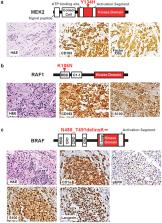
          Histiocytic neoplasms are a heterogeneous group of clonal hematopoietic disorders marked by diverse mutations in the mitogen-activated protein kinase (MAPK) pathway. 1, 2 For the 50% of histiocytosis patients with BRAFV600-mutations 35 , RAF inhibition is highly efficacious and has dramatically altered the natural history of the disease. 6, 7 Conversely, no standard therapy exists for the remaining 50% of patients lacking BRAFV600-mutations. While ERK dependence has been hypothesized to be a consistent feature across histiocytic neoplasms, this remains clinically unproven and many kinase mutations found in these patients have not been biologically characterized. We set out to evaluate ERK dependence in histiocytoses through a proof-of-concept clinical trial of the oral MEK1/2 inhibitor cobimetinib in patients with histiocytoses. Patients were enrolled regardless of tumor genotype. In parallel, novel MAPK alterations identified in treated patients were characterized for their ability to activate ERK. In 18 treated patients, the overall response rate (ORR) was 89% (90% CI: 73–100). Responses were durable, with no acquired resistance to date. At one year, 100% of responses were ongoing, and 94% of patients remained progression-free. Efficacy was observed regardless of genotype with responses achieved in patients with ARAF, BRAF, RAF1, NRAS, KRAS, MEK1, and MEK2 mutations. Consistent with observed responses, characterization of the novel mutations identified in treated patients confirmed them to be activating. Collectively, these data demonstrate that histiocytic neoplasms are characterized by remarkable dependence on MAPK signaling and, consequently, responsiveness to MEK inhibition. These results extend the benefits of molecularly targeted therapy to the entire spectrum of patients with histiocytosis.

          Related collections

          Most cited references22

          • Record: found
          • Abstract: found
          • Article: not found

          Combined vemurafenib and cobimetinib in BRAF-mutated melanoma.

          The combined inhibition of BRAF and MEK is hypothesized to improve clinical outcomes in patients with melanoma by preventing or delaying the onset of resistance observed with BRAF inhibitors alone. This randomized phase 3 study evaluated the combination of the BRAF inhibitor vemurafenib and the MEK inhibitor cobimetinib. We randomly assigned 495 patients with previously untreated unresectable locally advanced or metastatic BRAF V600 mutation-positive melanoma to receive vemurafenib and cobimetinib (combination group) or vemurafenib and placebo (control group). The primary end point was investigator-assessed progression-free survival. The median progression-free survival was 9.9 months in the combination group and 6.2 months in the control group (hazard ratio for death or disease progression, 0.51; 95% confidence interval [CI], 0.39 to 0.68; P<0.001). The rate of complete or partial response in the combination group was 68%, as compared with 45% in the control group (P<0.001), including rates of complete response of 10% in the combination group and 4% in the control group. Progression-free survival as assessed by independent review was similar to investigator-assessed progression-free survival. Interim analyses of overall survival showed 9-month survival rates of 81% (95% CI, 75 to 87) in the combination group and 73% (95% CI, 65 to 80) in the control group. Vemurafenib and cobimetinib was associated with a nonsignificantly higher incidence of adverse events of grade 3 or higher, as compared with vemurafenib and placebo (65% vs. 59%), and there was no significant difference in the rate of study-drug discontinuation. The number of secondary cutaneous cancers decreased with the combination therapy. The addition of cobimetinib to vemurafenib was associated with a significant improvement in progression-free survival among patients with BRAF V600-mutated metastatic melanoma, at the cost of some increase in toxicity. (Funded by F. Hoffmann-La Roche/Genentech; coBRIM ClinicalTrials.gov number, NCT01689519.).
            Bookmark
            • Record: found
            • Abstract: found
            • Article: not found

            Anchored multiplex PCR for targeted next-generation sequencing.

            We describe a rapid target enrichment method for next-generation sequencing, termed anchored multiplex PCR (AMP), that is compatible with low nucleic acid input from formalin-fixed paraffin-embedded (FFPE) specimens. AMP is effective in detecting gene rearrangements (without prior knowledge of the fusion partners), single nucleotide variants, insertions, deletions and copy number changes. Validation of a gene rearrangement panel using 319 FFPE samples showed 100% sensitivity (95% confidence limit: 96.5-100%) and 100% specificity (95% confidence limit: 99.3-100%) compared with reference assays. On the basis of our experience with performing AMP on 986 clinical FFPE samples, we show its potential as both a robust clinical assay and a powerful discovery tool, which we used to identify new therapeutically important gene fusions: ARHGEF2-NTRK1 and CHTOP-NTRK1 in glioblastoma, MSN-ROS1, TRIM4-BRAF, VAMP2-NRG1, TPM3-NTRK1 and RUFY2-RET in lung cancer, FGFR2-CREB5 in cholangiocarcinoma and PPL-NTRK1 in thyroid carcinoma. AMP is a scalable and efficient next-generation sequencing target enrichment method for research and clinical applications.
              Bookmark
              • Record: found
              • Abstract: found
              • Article: not found

              High prevalence of BRAF V600E mutations in Erdheim-Chester disease but not in other non-Langerhans cell histiocytoses.

              Histiocytoses are rare disorders of unknown origin with highly heterogeneous prognosis. BRAF mutations have been observed in Langerhans cell histiocytosis (LCH). We investigated the frequency of BRAF mutations in several types of histiocytoses. Histology from 127 patients with histiocytoses were reviewed. Detection of BRAF(V600) mutations was performed by pyrosequencing of DNA extracted from paraffin embedded samples. Diagnoses of Erdheim-Chester disease (ECD), LCH, Rosai-Dorfman disease, juvenile xanthogranuloma, histiocytic sarcoma, xanthoma disseminatum, interdigitating dendritic cell sarcoma, and necrobiotic xanthogranuloma were performed in 46, 39, 23, 12, 3, 2, 1, and 1 patients, respectively. BRAF status was obtained in 93 cases. BRAF(V600E) mutations were detected in 13 of 24 (54%) ECD, 11 of 29 (38%) LCH, and none of the other histiocytoses. Four patients with ECD died of disease. The high frequency of BRAF(V600E) in LCH and ECD suggests a common origin of these diseases. Treatment with vemurafenib should be investigated in patients with malignant BRAF(V600E) histiocytosis.
                Bookmark

                Author and article information

                Journal
                0410462
                6011
                Nature
                Nature
                Nature
                0028-0836
                1476-4687
                14 February 2019
                13 March 2019
                March 2019
                13 September 2019
                : 567
                : 7749
                : 521-524
                Affiliations
                [1 ]Dept. of Neurology, Memorial Sloan Kettering Cancer Center, New York, NY, 10065
                [2 ]Dept. of Pathology, Memorial Sloan Kettering Cancer Center, New York, NY, 10065
                [3 ]Human Oncology and Pathogenesis Program, Dept. of Medicine, Memorial Sloan Kettering Cancer Center, New York, NY 10065
                [4 ]Dept. of Radiology, Memorial Sloan Kettering Cancer Center, New York, NY, 10065
                [5 ]Dept. of Epidemiology and Statistics, Memorial Sloan Kettering Cancer Center, New York, NY, 10065
                [6 ]Ophthalmic Oncology Service, Dept. of Medicine, Memorial Sloan Kettering Cancer Center, New York, NY, 10065
                [7 ]Dept. of Medicine, Memorial Sloan Kettering Cancer Center, New York, NY, 10065
                [8 ]Dermatology Service, Dept. of Medicine, Memorial Sloan Kettering Cancer Center, New York, NY, 10065
                [9 ]Molecular Pharmacology and Chemistry Program, Memorial Sloan Kettering Cancer Center, New York, NY 10065
                [10 ]Weill Cornell Medical College, New York, NY, 10065
                [11 ]Laboratory of Pathology, National Cancer Institute, Bethesda, MD
                Author notes
                [*]

                These authors contributed equally to this work.

                Author contributions ELD, OAW, and DMH designed the study. MK, LB, and HC processed all patient material for isolation of DNA and RNA for genomic analyses. BHD cloned all cDNAs and performed in vitro studies of their effects on cell growth and signaling under the supervision of NR and OAW. BHD also analyzed genomic data under the supervision of OAW. JB collected data and samples for processing by MK, LB, and HC. ELD, JHF, RR, ML, and LAB enrolled and treated the patients. ED and AI performed statistical analyses. GAU and EJY evaluated all radiographic studies. ELD, BHD, NO, AD, OAW, and DMH analyzed the clinical and laboratory data. ELD, BHD, OAW, and DMH prepared the manuscript with help from all co-authors.

                [** ]Co-corresponding. Correspondence: Omar Abdel-Wahab, Memorial Sloan Kettering Cancer, 1275 York Avenue, New York, NY 10065, +16468883487, abdelwao@ 123456mskcc.org , David M. Hyman,, Memorial Sloan Kettering Cancer Center, 1275 York Avenue, New York, NY 10065, +16468884544, hymand@ 123456mskcc.org
                Article
                NIHMS1521555
                10.1038/s41586-019-1012-y
                6438729
                30867592
                4a734c5f-61a7-4afb-9d53-08af7b56624e

                Author Information Reprints and permission information is available at www.nature.com/reprints.

                Users may view, print, copy, and download text and data-mine the content in such documents, for the purposes of academic research, subject always to the full Conditions of use: http://www.nature.com/authors/editorial_policies/license.html#terms

                History
                Categories
                Article

                Uncategorized
                Uncategorized

                Comments

                Comment on this article