2
views
0
recommends
+1 Recommend
0 collections
    0
    shares
      • Record: found
      • Abstract: found
      • Article: found
      Is Open Access

      Nascent mutant Huntingtin exon 1 chains do not stall on ribosomes during translation but aggregates do recruit machinery involved in ribosome quality control and RNA

      research-article

      Read this article at

      Bookmark
          There is no author summary for this article yet. Authors can add summaries to their articles on ScienceOpen to make them more accessible to a non-specialist audience.

          Abstract

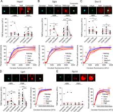
          Mutations that cause Huntington’s Disease involve a polyglutamine (polyQ) sequence expansion beyond 35 repeats in exon 1 of Huntingtin. Intracellular inclusion bodies of mutant Huntingtin protein are a key feature of Huntington’s disease brain pathology. We previously showed that in cell culture the formation of inclusions involved the assembly of disordered structures of mHtt exon 1 fragments (Httex1) and they were enriched with translational machinery when first formed. We hypothesized that nascent mutant Httex1 chains co-aggregate during translation by phase separation into liquid-like disordered aggregates and then convert to more rigid, amyloid structures. Here we further examined the mechanisms of inclusion assembly in a human epithelial kidney (AD293) cell culture model. We found mHttex1 did not appear to stall translation of its own nascent chain, or at best was marginal. We also found the inclusions appeared to recruit low levels of RNA but there was no difference in enrichment between early formed and mature inclusions. Proteins involved in translation or ribosome quality control were co-recruited to the inclusions (Ltn1 Rack1) compared to a protein not anticipated to be involved (NACAD), but there was no major specificity of enrichment in the early formed inclusions compared to mature inclusions. Furthermore, we observed co-aggregation with other proteins previously identified in inclusions, including Upf1 and chaperone-like proteins Sgta and Hspb1, which also suppressed aggregation at high co-expression levels. The newly formed inclusions also contained immobile mHttex1 molecules which points to the disordered aggregates being mechanically rigid prior to amyloid formation. Collectively our findings show little evidence that inclusion assembly arises by a discrete clustering of stalled nascent chains and associated quality control machinery. Instead, the machinery appear to be recruited continuously, or secondarily, to the nucleation of inclusion formation.

          Related collections

          Most cited references26

          • Record: found
          • Abstract: found
          • Article: not found

          Huntingtin-encoded polyglutamine expansions form amyloid-like protein aggregates in vitro and in vivo.

          The mechanism by which an elongated polyglutamine sequence causes neurodegeneration in Huntington's disease (HD) is unknown. In this study, we show that the proteolytic cleavage of a GST-huntingtin fusion protein leads to the formation of insoluble high molecular weight protein aggregates only when the polyglutamine expansion is in the pathogenic range. Electron micrographs of these aggregates revealed a fibrillar or ribbon-like morphology, reminiscent of scrapie prions and beta-amyloid fibrils in Alzheimer's disease. Subcellular fractionation and ultrastructural techniques showed the in vivo presence of these structures in the brains of mice transgenic for the HD mutation. Our in vitro model will aid in an eventual understanding of the molecular pathology of HD and the development of preventative strategies.
            Bookmark
            • Record: found
            • Abstract: found
            • Article: not found

            Towards a transgenic model of Huntington's disease in a non-human primate.

            Non-human primates are valuable for modelling human disorders and for developing therapeutic strategies; however, little work has been reported in establishing transgenic non-human primate models of human diseases. Huntington's disease (HD) is an autosomal dominant neurodegenerative disorder characterized by motor impairment, cognitive deterioration and psychiatric disturbances followed by death within 10-15 years of the onset of the symptoms. HD is caused by the expansion of cytosine-adenine-guanine (CAG, translated into glutamine) trinucleotide repeats in the first exon of the human huntingtin (HTT) gene. Mutant HTT with expanded polyglutamine (polyQ) is widely expressed in the brain and peripheral tissues, but causes selective neurodegeneration that is most prominent in the striatum and cortex of the brain. Although rodent models of HD have been developed, these models do not satisfactorily parallel the brain changes and behavioural features observed in HD patients. Because of the close physiological, neurological and genetic similarities between humans and higher primates, monkeys can serve as very useful models for understanding human physiology and diseases. Here we report our progress in developing a transgenic model of HD in a rhesus macaque that expresses polyglutamine-expanded HTT. Hallmark features of HD, including nuclear inclusions and neuropil aggregates, were observed in the brains of the HD transgenic monkeys. Additionally, the transgenic monkeys showed important clinical features of HD, including dystonia and chorea. A transgenic HD monkey model may open the way to understanding the underlying biology of HD better, and to the development of potential therapies. Moreover, our data suggest that it will be feasible to generate valuable non-human primate models of HD and possibly other human genetic diseases.
              Bookmark
              • Record: found
              • Abstract: not found
              • Article: not found

              Analysis of the aphthovirus 2A/2B polyprotein ‘cleavage’ mechanism indicates not a proteolytic reaction, but a novel translational effect: a putative ribosomal ‘skip’

                Bookmark

                Author and article information

                Contributors
                Role: Data curationRole: Formal analysisRole: InvestigationRole: MethodologyRole: Writing – review & editing
                Role: Formal analysisRole: InvestigationRole: SoftwareRole: Writing – review & editing
                Role: InvestigationRole: Methodology
                Role: Formal analysisRole: InvestigationRole: Methodology
                Role: Formal analysisRole: MethodologyRole: Supervision
                Role: ConceptualizationRole: Formal analysisRole: Funding acquisitionRole: Project administrationRole: ResourcesRole: SupervisionRole: Writing – original draftRole: Writing – review & editing
                Role: Editor
                Journal
                PLoS One
                PLoS ONE
                plos
                plosone
                PLoS ONE
                Public Library of Science (San Francisco, CA USA )
                1932-6203
                31 July 2020
                2020
                : 15
                : 7
                : e0233583
                Affiliations
                [001]Department of Biochemistry and Molecular Biology; and Bio21 Molecular Science and Biotechnology Institute, The University of Melbourne, VIC, Australia
                University of Florida, UNITED STATES
                Author notes

                Competing Interests: The authors have declared that no competing interests exist.

                Author information
                http://orcid.org/0000-0002-5345-8360
                http://orcid.org/0000-0002-9965-2847
                Article
                PONE-D-20-13286
                10.1371/journal.pone.0233583
                7394408
                32735619
                4c82c8e6-391e-4d9e-8b07-622fe4d12682
                © 2020 Ormsby et al

                This is an open access article distributed under the terms of the Creative Commons Attribution License, which permits unrestricted use, distribution, and reproduction in any medium, provided the original author and source are credited.

                History
                : 6 May 2020
                : 13 July 2020
                Page count
                Figures: 5, Tables: 1, Pages: 17
                Funding
                Funded by: funder-id http://dx.doi.org/10.13039/501100000925, National Health and Medical Research Council;
                Award ID: APP1184166
                Award Recipient :
                Funded by: funder-id http://dx.doi.org/10.13039/501100000925, National Health and Medical Research Council;
                Award ID: APP1161803
                Award Recipient :
                Funded by: funder-id http://dx.doi.org/10.13039/501100000925, National Health and Medical Research Council;
                Award ID: APP1102059
                Award Recipient :
                Funded by: funder-id http://dx.doi.org/10.13039/501100000925, National Health and Medical Research Council;
                Award ID: APP1154352
                Award Recipient :
                This work was funded by grants and fellowships from the National Health and Medical Research Council Project to D.M.H. (APP1184166, APP1161803, APP1102059, APP1154352). The funders had no role in study design, data collection and analysis, decision to publish, or preparation of the manuscript.
                Categories
                Research Article
                Biology and Life Sciences
                Biochemistry
                Ribosomes
                Biology and Life Sciences
                Cell Biology
                Cellular Structures and Organelles
                Ribosomes
                Research and Analysis Methods
                Specimen Preparation and Treatment
                Staining
                Cell Staining
                Engineering and Technology
                Industrial Engineering
                Quality Control
                Biology and Life Sciences
                Cell Biology
                Cytoplasmic Inclusions
                Research and Analysis Methods
                Spectrum Analysis Techniques
                Spectrophotometry
                Cytophotometry
                Flow Cytometry
                Research and Analysis Methods
                Microscopy
                Electron Microscopy
                Scanning Electron Microscopy
                Biology and Life Sciences
                Genetics
                Gene Expression
                Protein Translation
                Biology and Life Sciences
                Molecular Biology
                Molecular Biology Techniques
                Transfection
                Research and Analysis Methods
                Molecular Biology Techniques
                Transfection
                Custom metadata
                All relevant data are within the paper and its Supporting Information files.

                Uncategorized
                Uncategorized

                Comments

                Comment on this article