Search for authorsSearch for similar articles
7
views
0
recommends
+1 Recommend
0 collections
    0
    shares
      • Record: found
      • Abstract: found
      • Article: found
      Is Open Access

      The architecture of functional lateralisation and its relationship to callosal connectivity in the human brain

      research-article

      Read this article at

      Bookmark
          There is no author summary for this article yet. Authors can add summaries to their articles on ScienceOpen to make them more accessible to a non-specialist audience.

          Abstract

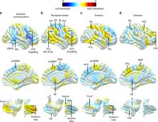
          Functional lateralisation is a fundamental principle of the human brain. However, a comprehensive taxonomy of functional lateralisation and its organisation in the brain is missing. Here, we report the first complete map of functional hemispheric asymmetries in the human brain, reveal its low dimensional structure, and its relationship with structural inter-hemispheric connectivity. Our results suggest that the lateralisation of brain functions is distributed along four functional axes: symbolic communication, perception/action, emotion, and decision-making. The similarity between this finding and recent work on neurological symptoms give rise to new hypotheses on the mechanisms that support brain recovery after a brain lesion. We also report that cortical regions showing asymmetries in task-evoked activity have reduced connections with the opposite hemisphere. This latter result suggests that during evolution, brain size expansion led to functional lateralisation to avoid excessive conduction delays between the hemispheres.

          Abstract

          Many functions of the human brain are lateralised i.e. associated more strongly with either the left or the right hemisphere of the brain. Here, the authors report the first complete map of functional asymmetries in the human brain, and its relationship with structural inter-hemispheric connectivity.

          Related collections

          Most cited references46

          • Record: found
          • Abstract: found
          • Article: not found

          The Fusiform Face Area: A Module in Human Extrastriate Cortex Specialized for Face Perception

          Using functional magnetic resonance imaging (fMRI), we found an area in the fusiform gyrus in 12 of the 15 subjects tested that was significantly more active when the subjects viewed faces than when they viewed assorted common objects. This face activation was used to define a specific region of interest individually for each subject, within which several new tests of face specificity were run. In each of five subjects tested, the predefined candidate “face area” also responded significantly more strongly to passive viewing of (1) intact than scrambled two-tone faces, (2) full front-view face photos than front-view photos of houses, and (in a different set of five subjects) (3) three-quarter-view face photos (with hair concealed) than photos of human hands; it also responded more strongly during (4) a consecutive matching task performed on three-quarter-view faces versus hands. Our technique of running multiple tests applied to the same region defined functionally within individual subjects provides a solution to two common problems in functional imaging: (1) the requirement to correct for multiple statistical comparisons and (2) the inevitable ambiguity in the interpretation of any study in which only two or three conditions are compared. Our data allow us to reject alternative accounts of the function of the fusiform face area (area “FF”) that appeal to visual attention, subordinate-level classification, or general processing of any animate or human forms, demonstrating that this region is selectively involved in the perception of faces.
            Bookmark
            • Record: found
            • Abstract: found
            • Article: not found

            Multiband multislice GE-EPI at 7 tesla, with 16-fold acceleration using partial parallel imaging with application to high spatial and temporal whole-brain fMRI.

            Parallel imaging in the form of multiband radiofrequency excitation, together with reduced k-space coverage in the phase-encode direction, was applied to human gradient echo functional MRI at 7 T for increased volumetric coverage and concurrent high spatial and temporal resolution. Echo planar imaging with simultaneous acquisition of four coronal slices separated by 44mm and simultaneous 4-fold phase-encoding undersampling, resulting in 16-fold acceleration and up to 16-fold maximal aliasing, was investigated. Task/stimulus-induced signal changes and temporal signal behavior under basal conditions were comparable for multiband and standard single-band excitation and longer pulse repetition times. Robust, whole-brain functional mapping at 7 T, with 2 x 2 x 2mm(3) (pulse repetition time 1.25 sec) and 1 x 1 x 2mm(3) (pulse repetition time 1.5 sec) resolutions, covering fields of view of 256 x 256 x 176 mm(3) and 192 x 172 x 176 mm(3), respectively, was demonstrated with current gradient performance. (c) 2010 Wiley-Liss, Inc.
              Bookmark
              • Record: found
              • Abstract: not found
              • Article: not found

              Mapping brain asymmetry.

                Bookmark

                Author and article information

                Contributors
                vyacheslav.karolis@ndcn.ox.ac.uk
                michel.thiebaut@gmail.com
                Journal
                Nat Commun
                Nat Commun
                Nature Communications
                Nature Publishing Group UK (London )
                2041-1723
                29 March 2019
                29 March 2019
                2019
                : 10
                : 1417
                Affiliations
                [1 ]ISNI 0000 0001 2308 1657, GRID grid.462844.8, Brain Connectivity and Behaviour Laboratory, , Sorbonne Universities, ; Paris, France
                [2 ]ISNI 0000 0004 0620 5939, GRID grid.425274.2, Frontlab, , Institut du Cerveau et de la Moelle épinière (ICM), ; Sorbonne Universities, Inserm U 1127, CNRS UMR 7225, Paris, France
                [3 ]ISNI 0000 0004 1757 3470, GRID grid.5608.b, Department of Neuroscience, , University of Padova, ; Padova, Italy
                [4 ]ISNI 0000 0004 1757 3470, GRID grid.5608.b, Padova Neuroscience Center (PNC), , University of Padova, ; Padova, Italy
                [5 ]Venetian Institute of Molecular Medicine, Fondazione Biomedica, Padova, Italy
                [6 ]ISNI 0000 0001 2355 7002, GRID grid.4367.6, Department of Neurology, , Washington University, ; Saint Louis, MO USA
                [7 ]ISNI 0000 0001 2355 7002, GRID grid.4367.6, Department of Radiology, , Washington University, ; Saint Louis, MO USA
                [8 ]ISNI 0000 0001 2355 7002, GRID grid.4367.6, Department of Neuroscience, , Washington University, ; Saint Louis, MO USA
                [9 ]ISNI 0000 0001 2106 639X, GRID grid.412041.2, Groupe d’Imagerie Neurofonctionnelle, Institut des Maladies Neurodégénératives-UMR 5293, CNRS, , CEA University of Bordeaux, ; Bordeaux, France
                Author information
                http://orcid.org/0000-0003-2074-6910
                http://orcid.org/0000-0002-0329-1814
                Article
                9344
                10.1038/s41467-019-09344-1
                6441088
                30926845
                4d9a9d03-d7c6-41f1-b55e-14fb537d3b63
                © The Author(s) 2019

                Open Access This article is licensed under a Creative Commons Attribution 4.0 International License, which permits use, sharing, adaptation, distribution and reproduction in any medium or format, as long as you give appropriate credit to the original author(s) and the source, provide a link to the Creative Commons license, and indicate if changes were made. The images or other third party material in this article are included in the article’s Creative Commons license, unless indicated otherwise in a credit line to the material. If material is not included in the article’s Creative Commons license and your intended use is not permitted by statutory regulation or exceeds the permitted use, you will need to obtain permission directly from the copyright holder. To view a copy of this license, visit http://creativecommons.org/licenses/by/4.0/.

                History
                : 23 July 2018
                : 5 March 2019
                Categories
                Article
                Custom metadata
                © The Author(s) 2019

                Uncategorized
                Uncategorized

                Comments

                Comment on this article