3
views
0
recommends
+1 Recommend
0 collections
    0
    shares
      • Record: found
      • Abstract: found
      • Article: found
      Is Open Access

      CT‐based Morphometric Analysis of Approach of Percutaneous Transforaminal Endoscopic Lumbar Interbody Fusion

      research-article

      Read this article at

      Bookmark
          There is no author summary for this article yet. Authors can add summaries to their articles on ScienceOpen to make them more accessible to a non-specialist audience.

          Abstract

          Objectives

          A radiographic study was designed to measure the relationship of the exiting nerve root and its surroundings to the corresponding intervertebral disc for percutaneous transforaminal endoscopic lumbar interbody fusion to better understand the regional anatomy and to improve clinical applications.

          Methods

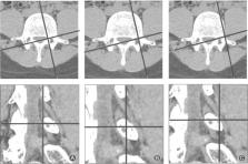
          A retrospective study from January 2017 to October 2017 was conducted at Tianjin Hospital. CT images were obtained from patients presenting low back pain (110 patients), and analysis was performed bilaterally from L 2‐3 to L 5S 1. In the rotating coronal plane we analyzed: the nerve root–dural sac distance at the superior and inferior margins of the disc (Js, Ji); the nerve root–pedicle distance at the medial, middle, and lateral borders of the pedicle (Pa, Pb, Pc); the pedicle width (W); and the safe working zone, defined as a trapezoid bounded by the inferior pedicle and the exiting nerve root (S). In the transverse plane, the nerve root‐articular process and the shortest distance for the nerve root‐articular process joint surface were analyzed at the superior and inferior margins of the disc (Gs, Gi), respectively. The groups were analyzed using ANOVA, and paired t‐tests were used to compare the left and right sides.

          Results

          From L 2‐3 to L 5S 1, the distance of the nerve root to the dural sac was larger at the inferior margin of the disc. From L 2‐3 to L 5S 1, each segment of the vertebral nerve root‐pedicle distance gradually decreased from medial to lateral. From L 2‐3 to L 5S 1, the distance from the exiting nerve root to the middle and lateral margins of the pedicle gradually decreased, with L 5S 1 being the minimum. Some significant differences were observed between the left and right sides for L 4‐5 and L 5S 1. The pedicle width of the vertebral body and the mean area for the safe working zone gradually increased from L 2‐3 to L 5S 1. In the axial plane, the shortest distance between the nerve root and articular process joint surface at the inferior margin of the disc was greater than the distance for the nerve root to the articular process at the superior margin of the disc from L 2‐3 to L 5S 1. There were no significant differences between the two sides.

          Conclusions

          It is more difficult to implant a cage with a width of 10 mm above the L 3‐4 level. By removing part of the superior articular process, the safe working area can be expanded, and damage to the nerve or other structures can be avoided when implanting a cage.

          Related collections

          Most cited references24

          • Record: found
          • Abstract: found
          • Article: found
          Is Open Access

          Complications and Morbidities of Mini-open Anterior Retroperitoneal Lumbar Interbody Fusion: Oblique Lumbar Interbody Fusion in 179 Patients

          Study Design A retrospective study including 179 patients who underwent oblique lumbar interbody fusion (OLIF) at one institution. Purpose To report the complications associated with a minimally invasive technique of a retroperitoneal anterolateral approach to the lumbar spine. Overview of Literature Different approaches to the lumbar spine have been proposed, but they are associated with an increased risk of complications and a longer operation. Methods A total of 179 patients with previous posterior instrumented fusion undergoing OLIF were included. The technique is described in terms of: the number of levels fused, operative time and blood loss. Persurgical and postsurgical complications were noted. Results Patients were age 54.1 ± 10.6 with a BMI of 24.8 ± 4.1 kg/m2. The procedure was performed in the lumbar spine at L1-L2 in 4, L2-L3 in 54, L3-L4 in 120, L4-L5 in 134, and L5-S1 in 6 patients. It was done at 1 level in 56, 2 levels in 107, and 3 levels in 16 patients. Surgery time and blood loss were, respectively, 32.5 ± 13.2 minutes and 57 ± 131 ml per level fused. There were 19 patients with a single complication and one with two complications, including two patients with postoperative radiculopathy after L3-5 OLIF. There was no abdominal weakness or herniation. Conclusions Minimally invasive OLIF can be performed easily and safely in the lumbar spine from L2 to L5, and at L1-2 for selected cases. Up to 3 levels can be addressed through a 'sliding window'. It is associated with minimal blood loss and short operations, and with decreased risk of abdominal wall weakness or herniation.
            Bookmark
            • Record: found
            • Abstract: found
            • Article: not found

            [A one-stager procedure in operative treatment of spondylolistheses: dorsal traction-reposition and anterior fusion (author's transl)].

            On account of 41 cases of spondylolisthesis a one-stage operation for repositioning and stabilisation using Harrington's instrumentation with anterior intercorporal spine fusion is recommended. The advantage of this procedure is a secure fusion and a short hospitalisation. After 6 months the patient usually is rehabilitated.
              Bookmark
              • Record: found
              • Abstract: not found
              • Article: not found

              The treatment of ruptured lumbar intervertebral discs by vertebral body fusion. I. Indications, operative technique, after care.

              R Cloward (1953)
                Bookmark

                Author and article information

                Contributors
                xubaoshan99@126.com
                Journal
                Orthop Surg
                Orthop Surg
                10.1111/(ISSN)1757-7861
                OS
                Orthopaedic Surgery
                John Wiley & Sons Australia, Ltd (Melbourne )
                1757-7853
                1757-7861
                21 March 2019
                April 2019
                : 11
                : 2 ( doiID: 10.1111/os.2019.11.issue-2 )
                : 212-220
                Affiliations
                [ 1 ] Graduate School of Tianjin Medical University Tianjin China
                [ 2 ] Department of Minimally Invasive Spine Surgery Tianjin Hospital Tianjin China
                [ 3 ] Department of Radiology Tianjin Hospital Tianjin China
                Author notes
                [*] [* ] Address for correspondence Bao‐shan Xu, MD, PhD, Department of Minimally Invasive Spine Surgery, Tianjin Hospital, 406 Jie Fang Nan Road, Hexi District, Tianjin, China 300211 Email: xubaoshan99@ 123456126.com
                [†]

                Both authors contributed equally to this project and manuscript preparation.

                Author information
                https://orcid.org/0000-0002-2447-7562
                Article
                OS12434
                10.1111/os.12434
                6594482
                30895721
                4e02e823-5e7a-43e5-9fef-b4136d7ad197
                © 2019 The Authors. Orthopaedic Surgery published by Chinese Orthopaedic Association and John Wiley & Sons Australia, Ltd

                This is an open access article under the terms of the http://creativecommons.org/licenses/by/4.0/ License, which permits use, distribution and reproduction in any medium, provided the original work is properly cited.

                History
                : 20 December 2018
                : 21 January 2019
                : 21 January 2019
                Page count
                Figures: 5, Tables: 3, Pages: 9, Words: 6807
                Funding
                Funded by: National Natural Science Foundation of China
                Award ID: 31670983
                Award ID: 31500781
                Funded by: Natural Science Foundation of Tianjin
                Award ID: 15JCYBJC25300
                Funded by: Key Research Foundation of the Tianjin Health Bureau
                Award ID: 14KG121
                Categories
                Clinical Article
                Clinical Articles
                Custom metadata
                2.0
                os12434
                April 2019
                Converter:WILEY_ML3GV2_TO_NLMPMC version:5.6.4 mode:remove_FC converted:10.06.2019

                computed tomography,endoscopy,interbody fusion cage,nerve root,spinal fusion

                Comments

                Comment on this article