64
views
0
recommends
+1 Recommend
0 collections
    0
    shares
      • Record: found
      • Abstract: found
      • Article: not found

      PINK1 Is Necessary for Long Term Survival and Mitochondrial Function in Human Dopaminergic Neurons

      research-article

      Read this article at

      ScienceOpenPublisherPMC
      Bookmark
          There is no author summary for this article yet. Authors can add summaries to their articles on ScienceOpen to make them more accessible to a non-specialist audience.

          Abstract

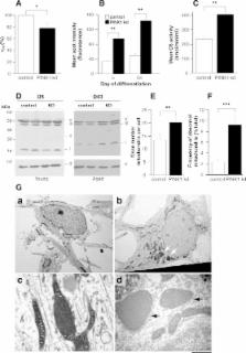
          Parkinson's disease (PD) is a common age-related neurodegenerative disease and it is critical to develop models which recapitulate the pathogenic process including the effect of the ageing process. Although the pathogenesis of sporadic PD is unknown, the identification of the mendelian genetic factor PINK1 has provided new mechanistic insights. In order to investigate the role of PINK1 in Parkinson's disease, we studied PINK1 loss of function in human and primary mouse neurons. Using RNAi, we created stable PINK1 knockdown in human dopaminergic neurons differentiated from foetal ventral mesencephalon stem cells, as well as in an immortalised human neuroblastoma cell line. We sought to validate our findings in primary neurons derived from a transgenic PINK1 knockout mouse. For the first time we demonstrate an age dependent neurodegenerative phenotype in human and mouse neurons. PINK1 deficiency leads to reduced long-term viability in human neurons, which die via the mitochondrial apoptosis pathway. Human neurons lacking PINK1 demonstrate features of marked oxidative stress with widespread mitochondrial dysfunction and abnormal mitochondrial morphology. We report that PINK1 plays a neuroprotective role in the mitochondria of mammalian neurons, especially against stress such as staurosporine. In addition we provide evidence that cellular compensatory mechanisms such as mitochondrial biogenesis and upregulation of lysosomal degradation pathways occur in PINK1 deficiency. The phenotypic effects of PINK1 loss-of-function described here in mammalian neurons provides mechanistic insight into the age-related degeneration of nigral dopaminergic neurons seen in PD.

          Related collections

          Most cited references49

          • Record: found
          • Abstract: found
          • Article: not found

          ROS, mitochondria and the regulation of autophagy.

          Accumulation of reactive oxygen species (ROS) is an oxidative stress to which cells respond by activating various defense mechanisms or, finally, by dying. At low levels, however, ROS act as signaling molecules in various intracellular processes. Autophagy, a process by which eukaryotic cells degrade and recycle macromolecules and organelles, has an important role in the cellular response to oxidative stress. Here, we review recent reports suggesting a regulatory role for ROS of mitochondrial origin as signaling molecules in autophagy, leading, under different circumstances, to either survival or cell death. We then discuss the relationship between mitochondria and autophagosomes and propose that mitochondria have an essential role in autophagosome biogenesis.
            Bookmark
            • Record: found
            • Abstract: found
            • Article: not found

            The PINK1/Parkin pathway regulates mitochondrial morphology.

            Loss-of-function mutations in the PTEN-induced kinase 1 (PINK1) or parkin genes, which encode a mitochondrially localized serine/threonine kinase and a ubiquitin-protein ligase, respectively, result in recessive familial forms of Parkinsonism. Genetic studies in Drosophila indicate that PINK1 acts upstream of Parkin in a common pathway that influences mitochondrial integrity in a subset of tissues, including flight muscle and dopaminergic neurons. The mechanism by which PINK1 and Parkin influence mitochondrial integrity is currently unknown, although mutations in the PINK1 and parkin genes result in enlarged or swollen mitochondria, suggesting a possible regulatory role for the PINK1/Parkin pathway in mitochondrial morphology. To address this hypothesis, we examined the influence of genetic alterations affecting the machinery that governs mitochondrial morphology on the PINK1 and parkin mutant phenotypes. We report that heterozygous loss-of-function mutations of drp1, which encodes a key mitochondrial fission-promoting component, are largely lethal in a PINK1 or parkin mutant background. Conversely, the flight muscle degeneration and mitochondrial morphological alterations that result from mutations in PINK1 and parkin are strongly suppressed by increased drp1 gene dosage and by heterozygous loss-of-function mutations affecting the mitochondrial fusion-promoting factors OPA1 and Mfn2. Finally, we find that an eye phenotype associated with increased PINK1/Parkin pathway activity is suppressed by perturbations that reduce mitochondrial fission and enhanced by perturbations that reduce mitochondrial fusion. Our studies suggest that the PINK1/Parkin pathway promotes mitochondrial fission and that the loss of mitochondrial and tissue integrity in PINK1 and parkin mutants derives from reduced mitochondrial fission.
              Bookmark
              • Record: found
              • Abstract: found
              • Article: not found

              Mitochondrial pathology and muscle and dopaminergic neuron degeneration caused by inactivation of Drosophila Pink1 is rescued by Parkin.

              Mutations in Pink1, a gene encoding a Ser/Thr kinase with a mitochondrial-targeting signal, are associated with Parkinson's disease (PD), the most common movement disorder characterized by selective loss of dopaminergic neurons. The mechanism by which loss of Pink1 leads to neurodegeneration is not understood. Here we show that inhibition of Drosophila Pink1 (dPink1) function results in energy depletion, shortened lifespan, and degeneration of select indirect flight muscles and dopaminergic neurons. The muscle pathology was preceded by mitochondrial enlargement and disintegration. These phenotypes could be rescued by the wild type but not the pathogenic C-terminal deleted form of human Pink1 (hPink1). The muscle and dopaminergic phenotypes associated with dPink1 inactivation show similarity to that seen in parkin mutant flies and could be suppressed by the overexpression of Parkin but not DJ-1. Consistent with the genetic rescue results, we find that, in dPink1 RNA interference (RNAi) animals, the level of Parkin protein is significantly reduced. Together, these results implicate Pink1 and Parkin in a common pathway that regulates mitochondrial physiology and cell survival in Drosophila.
                Bookmark

                Author and article information

                Contributors
                Role: Editor
                Journal
                PLoS ONE
                plos
                plosone
                PLoS ONE
                Public Library of Science (San Francisco, USA )
                1932-6203
                2008
                18 June 2008
                : 3
                : 6
                : e2455
                Affiliations
                [1 ]Department of Molecular Neuroscience, Institute of Neurology, London, United Kingdom
                [2 ]Medical Molecular Biology Unit, Institute of Child Health, London, United Kingdom
                [3 ]Department of Physiology, University College London, London, United Kingdom
                [4 ]ReNeuron Ltd, Guildford, United Kingdom
                [5 ]Eisai London Research Laboratories Ltd, London, United Kingdom
                [6 ]Neurometabolic Unit, National Hospital for Neurology and Neurosurgery, London, United Kingdom
                [7 ]Cancer Research United Kingdom, London, United Kingdom
                [8 ]Department of Neurodegenerative Disease, Institute of Neurology, London, United Kingdom
                [9 ]Birkbeck, University of London, London, United Kingdom
                University of Auckland, New Zealand
                Author notes

                Conceived and designed the experiments: JD ST NW SG LM PJ JT SH MD DL AW. Performed the experiments: SG ZY AA EM GK LS IH KK AW ED. Analyzed the data: ST SG AA LS IH SH AW. Contributed reagents/materials/analysis tools: JD ST EM GK LM PJ JT SH MD DL ED. Wrote the paper: SG AW.

                ¶ These authors also contributed equally to this work.

                Article
                08-PONE-RA-03295R1
                10.1371/journal.pone.0002455
                2413012
                18560593
                5068e700-528e-470a-be9c-c9601b9f38e2
                Wood-Kaczmar et al. This is an open-access article distributed under the terms of the Creative Commons Attribution License, which permits unrestricted use, distribution, and reproduction in any medium, provided the original author and source are credited.
                History
                : 14 January 2008
                : 23 April 2008
                Page count
                Pages: 16
                Categories
                Research Article
                Neuroscience/Neuronal and Glial Cell Biology
                Neuroscience/Neuronal Signaling Mechanisms
                Neurological Disorders/Movement Disorders
                Neurological Disorders/Neurogenetics

                Uncategorized
                Uncategorized

                Comments

                Comment on this article