Inviting an author to review:
Find an author and click ‘Invite to review selected article’ near their name.
Search for authorsSearch for similar articles
71
views
0
recommends
+1 Recommend
0 collections
    0
    shares
      • Record: found
      • Abstract: found
      • Article: found
      Is Open Access

      Mesenchymal stromal cells for cutaneous wound healing in a rabbit model: pre-clinical study applicable in the pediatric surgical setting

      research-article

      Read this article at

      Bookmark
          There is no author summary for this article yet. Authors can add summaries to their articles on ScienceOpen to make them more accessible to a non-specialist audience.

          Abstract

          Objective

          Mesenchymal stromal cells (MSCs) expanded in vitro have been proposed as a potential therapy for congenital or acquired skin defects in pediatrics. The aim of this pre-clinical study was to investigate the effects of intradermal injections of MSC in experimental cutaneous wound repair comparing allogeneic and autologous adipose stem cells (ASCs) and autologous bone marrow-mesenchymal stromal cells (BM-MSCs).

          Methods

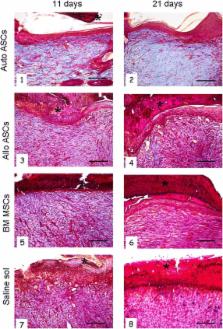
          Mesenchymal stromal cells were in vitro expanded from adipose and BM tissues of young female New Zealand rabbits. MSCs were characterized for plastic adhesion, surface markers, proliferation and differentiation capacity. When an adequate number of cells (ASCs 10 × 10 6 and BM-MSCs 3 × 10 6, because of their low rate of proliferation) was reached, two skin wounds were surgically induced in each animal. The first was topically treated with cell infusions, the second was used as a control. The intradermal inoculation included autologous or allogeneic ASCs or autologous BM-MSCs. For histological examination, animals were sacrificed and wounds were harvested after 11 and 21 days of treatment.

          Results

          Rabbit ASCs were isolated and expanded in vitro with relative abundance, cells expressed typical surface markers (CD49e, CD90 and CD29). Topically, ASC inoculation provided more rapid wound healing than BM-MSCs and controls. Improved re-epithelization, reduced inflammatory infiltration and increased collagen deposition were observed in biopsies from wounds treated with ASCs, with the best result in the autologous setting. ASCs also improved restoration of skin architecture during wound healing.

          Conclusion

          The use of ASCs may offer a promising solution to treat extended wounds. Pre-clinical studies are however necessary to validate the best skin regeneration technique, which could be used in pediatric surgical translational research.

          Related collections

          Most cited references37

          • Record: found
          • Abstract: found
          • Article: not found

          Concise review: mesenchymal stem/multipotent stromal cells: the state of transdifferentiation and modes of tissue repair--current views.

          Mesenchymal stem cells or multipotent stromal cells (MSCs) isolated from the bone marrow of adult organisms were initially characterized as plastic adherent, fibroblastoid cells with the capacity to generate heterotopic osseous tissue when transplanted in vivo. In recent years, MSCs or MSC-like cells have been shown to reside within the connective tissue of most organs, and their surface phenotype has been well described. A large number of reports have also indicated that the cells possess the capacity to transdifferentiate into epithelial cells and lineages derived from the neuroectoderm. The broad developmental plasticity of MSCs was originally thought to contribute to their demonstrated efficacy in a wide variety of experimental animal models of disease as well as in human clinical trials. However, new findings suggest that the ability of MSCs to alter the tissue microenvironment via secretion of soluble factors may contribute more significantly than their capacity for transdifferentiation in tissue repair. Herein, we critically evaluate the literature describing the plasticity of MSCs and offer insight into how the molecular and functional heterogeneity of this cell population, which reflects the complexity of marrow stroma as an organ system, may confound interpretation of their transdifferentiation potential. Additionally, we argue that this heterogeneity also provides a basis for the broad therapeutic efficacy of MSCs.
            Bookmark
            • Record: found
            • Abstract: found
            • Article: not found

            Wound healing: an overview of acute, fibrotic and delayed healing.

            Acute wounds normally heal in a very orderly and efficient manner characterized by four distinct, but overlapping phases: hemostasis, inflammation, proliferation and remodeling. Specific biological markers characterize healing of acute wounds. Likewise, unique biologic markers also characterize pathologic responses resulting in fibrosis and chronic non-healing ulcers. This review describes the major biological processes associated with both normal and pathologic healing. The normal healing response begins the moment the tissue is injured. As the blood components spill into the site of injury, the platelets come into contact with exposed collagen and other elements of the extracellular matrix. This contact triggers the platelets to release clotting factors as well as essential growth factors and cytokines such as platelet-derived growth factor (PDGF) and transforming growth factor beta (TGF-beta). Following hemostasis, the neutrophils then enter the wound site and begin the critical task of phagocytosis to remove foreign materials, bacteria and damaged tissue. As part of this inflammatory phase, the macrophages appear and continue the process of phagocytosis as well as releasing more PDGF and TGF beta. Once the wound site is cleaned out, fibroblasts migrate in to begin the proliferative phase and deposit new extracellular matrix. The new collagen matrix then becomes cross-linked and organized during the final remodeling phase. In order for this efficient and highly controlled repair process to take place, there are numerous cell-signaling events that are required. In pathologic conditions such as non-healing pressure ulcers, this efficient and orderly process is lost and the ulcers are locked into a state of chronic inflammation characterized by abundant neutrophil infiltration with associated reactive oxygen species and destructive enzymes. Healing proceeds only after the inflammation is controlled. On the opposite end of the spectrum, fibrosis is characterized by excessive matrix deposition and reduced remodeling. Often fibrotic lesions are associated with increased densities of mast cells. By understanding the functional relationships of these biological processes of normal compared to abnormal wound healing, hopefully new strategies can be designed to treat the pathological conditions.
              Bookmark
              • Record: found
              • Abstract: found
              • Article: not found

              Regulation of wound healing by growth factors and cytokines.

              Cutaneous wound healing is a complex process involving blood clotting, inflammation, new tissue formation, and finally tissue remodeling. It is well described at the histological level, but the genes that regulate skin repair have only partially been identified. Many experimental and clinical studies have demonstrated varied, but in most cases beneficial, effects of exogenous growth factors on the healing process. However, the roles played by endogenous growth factors have remained largely unclear. Initial approaches at addressing this question focused on the expression analysis of various growth factors, cytokines, and their receptors in different wound models, with first functional data being obtained by applying neutralizing antibodies to wounds. During the past few years, the availability of genetically modified mice has allowed elucidation of the function of various genes in the healing process, and these studies have shed light onto the role of growth factors, cytokines, and their downstream effectors in wound repair. This review summarizes the results of expression studies that have been performed in rodents, pigs, and humans to localize growth factors and their receptors in skin wounds. Most importantly, we also report on genetic studies addressing the functions of endogenous growth factors in the wound repair process.
                Bookmark

                Author and article information

                Contributors
                g.pelizzo@smatteo.pv.it
                ma.avanzini@smatteo.pv.it
                antonia.icaro@unipv.it
                hostim@hotmail.com
                p.romano@smatteo.pv.it
                l.avolio@smatteo.pv.it
                r.maccario@smatteo.pv.it
                massimo.dominici@unimore.it
                a.desilvestri@smatteo.pv.it
                erika.andreatta87@gmail.com
                federico.costanzo01@gmail.com
                melissamantelli@tiscali.it
                daniela.ingo31@libero.it
                mariaserena.piccinno@unimore.it
                v.calcaterra@smatteo.pv.it
                Journal
                J Transl Med
                J Transl Med
                Journal of Translational Medicine
                BioMed Central (London )
                1479-5876
                8 July 2015
                8 July 2015
                2015
                : 13
                : 219
                Affiliations
                [ ]Pediatric Surgery Unit, Fondazione IRCCS Policlinico San Matteo and University of Pavia, 27100 Pavia, Italy
                [ ]Immunology and Transplantation Laboratory/Cell Factory/Pediatric Hematology/Oncology Department, Fondazione IRCCS Policlinico San Matteo, Pavia, Italy
                [ ]Histology and Embryology Unit, Department of Public Health, Experimental Medicine and Forensic, University of Pavia, Pavia, Italy
                [ ]Laboratory of Cellular Therapies, Department of Medical and Surgical Sciences for Children and Adults, University Hospital of Modena and Reggio, Emilia, Italy
                [ ]Biometry and Clinical Epidemiology Unit, Scientific Direction, Fondazione IRCCS Policlinico San Matteo, Pavia, Italy
                [ ]Pediatric Unit, Fondazione IRCCS Policlinico San Matteo and University of Pavia, Pavia, Italy
                Article
                580
                10.1186/s12967-015-0580-3
                4495634
                26152232
                52b86a68-b1b1-4776-8823-5d9730ebc945
                © Pelizzo et al. 2015

                Open AccessThis article is distributed under the terms of the Creative Commons Attribution 4.0 International License ( http://creativecommons.org/licenses/by/4.0/), which permits unrestricted use, distribution, and reproduction in any medium, provided you give appropriate credit to the original author(s) and the source, provide a link to the Creative Commons license, and indicate if changes were made. The Creative Commons Public Domain Dedication waiver ( http://creativecommons.org/publicdomain/zero/1.0/) applies to the data made available in this article, unless otherwise stated.

                History
                : 11 May 2015
                : 24 June 2015
                Categories
                Research
                Custom metadata
                © The Author(s) 2015

                Medicine
                mesenchymal stromal cells,cutaneous wounds,adipose,bone marrow,pediatric surgery,regenerative medicine

                Comments

                Comment on this article