20
views
0
recommends
+1 Recommend
1 collections
    0
    shares
      • Record: found
      • Abstract: found
      • Article: found
      Is Open Access

      A molecular epidemiological survey of Babesia, Hepatozoon, Ehrlichia and Anaplasma infections of dogs in Japan

      research-article

      Read this article at

      Bookmark
          There is no author summary for this article yet. Authors can add summaries to their articles on ScienceOpen to make them more accessible to a non-specialist audience.

          Abstract

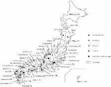
          Tick-borne diseases are often encountered in canine clinical practice. In the present study, a molecular epidemiological survey of dogs in Japan was conducted to understand the prevalence and geographical distribution of Babesia spp., Hepatozoon spp., Ehrlichia spp. and Anaplasma spp. Pathogen-derived DNA in blood samples obtained from 722 dogs with a history of exposure to ticks and/or fleas was examined by PCR. The prevalence of Babesia gibsoni, Babesia odocoilei-like species, Hepatozoon canis and Ehrlichia spp./ Anaplasma spp. was 2.4% (16/722), 0.1% (1/722), 2.5% (18/722) and 1.5% (11/722), respectively. While B. gibsoni and Ehrlichia spp./ Anaplasma spp. were detected in the western part of Japan, H. canis was detected in Tohoku area in addition to western and central parts of Japan.

          Related collections

          Most cited references18

          • Record: found
          • Abstract: found
          • Article: not found

          Ehrlichiosis and anaplasmosis in dogs and cats.

          In the time since canine ehrlichiosis due to Ehrlichia canis was first described in 1935 and first recognized in the United States in 1962, many key advances have been made in our understanding of the diversity of the rickettsial organisms responsible for ehrlichiosis and anaplasmosis in dogs and, occasionally, cats, the vectors capable of transmitting these agents, and the role these organisms play as both important veterinary pathogens and zoonotic disease agents. Despite considerable progress in the field, much remains to be learned regarding mechanisms contributing to pathogenesis, effective treatment modalities, and prevention strategies that best protect pet health. This article highlights current understanding of the transmission, diagnosis, and management of ehrlichiosis and anaplasmosis in dogs and cats. Copyright © 2010 Elsevier Inc. All rights reserved.
            Bookmark
            • Record: found
            • Abstract: found
            • Article: not found

            New advances in molecular epizootiology of canine hematic protozoa from Venezuela, Thailand and Spain.

            The prevalence of hematozoan infections (Hepatozoon canis and Babesia sp., particularly Babesia canis vogeli) in canids from Venezuela, Thailand and Spain was studied by amplification and sequencing of the 18S rRNA gene. H. canis infections caused simultaneously by two different isolates were confirmed by RFLP analysis in samples from all the geographic regions studied. In Venezuela, blood samples from 134 dogs were surveyed. Babesia infections were found in 2.24% of the dogs. Comparison of sequences of the 18S rRNA gene indicated that protozoan isolates were genetically identical to B. canis vogeli from Japan and Brazil. H. canis infected 44.77 per cent of the dogs. A representative sample of Venezuelan H. canis isolates (21.6% of PCR-positives) was sequenced. Many of them showed 18S rRNA gene sequences identical to H. canis Spain 2, albeit two less frequent genotypes were found in the sample studied. In Thailand, 20 dogs were analyzed. No infections caused by Babesia were diagnosed, whereas 30 per cent of the dogs were positive to hematozoan infection. Two protozoa isolates showing 99.7-100% identity to H. canis Spain 2 were found. In Spain, 250 dogs were studied. B. canis vogeli infected 0.01% of the animals. The sequence of the 18S rRNA gene in Spanish isolates of this protozoa was closely related to those previously deposited in GenBank (> 99% identity). Finally, 20 red foxes were screened for hematozoans employing semi-nested PCR and primers designed to detect Babesia/Theileria. Fifty percent of the foxes were positive to Theileria annae. In addition, it was found that the PCR assay was able as well to detect Hepatozoon infections. Thirty five percent of the foxes were infected with two different H. canis isolates showing 99.8-100% identity to Curupira 1 from Brazil.
              Bookmark
              • Record: found
              • Abstract: found
              • Article: not found

              Babesia odocoilei infection in a Saskatchewan elk (Cervus elaphus canadensis) herd.

              An 8-year-old female elk (Cervus elaphus canadensis) cow, presented for chronic severe weight loss and unthriftiness, was diagnosed with Babesia odocoilei infection based on blood smear evaluation, polymerase chain reaction (PCR), and DNA sequence analysis. Subsequently, velvet antler from a male that died acutely on the same farm was also PCR positive for Babesia spp. Both animals originated from a game ranch of Saskatchewan-bred and -raised animals with no known history of tick exposure, but with a history of numerous sudden deaths of unknown etiology. The presence of B. odocoilei in Canada might be a result of a recent introduction that could have deleterious effects on local wild ungulates or may represent discovery of a previously unrecognized endemic disease in local wildlife.
                Bookmark

                Author and article information

                Journal
                J Vet Med Sci
                J. Vet. Med. Sci
                JVMS
                The Journal of Veterinary Medical Science
                The Japanese Society of Veterinary Science
                0916-7250
                1347-7439
                01 May 2015
                October 2015
                : 77
                : 10
                : 1275-1279
                Affiliations
                [1) ]Laboratory of Small Animal Internal Medicine, Joint Faculty of Veterinary Medicine, Kagoshima University, 1–21–24 Korimoto, Kagoshima 890–0065, Japan
                [2) ]The United Graduate School of Veterinary Science, Yamaguchi University, 1677–1 Yoshida, Yamaguchi 753–8511, Japan
                [3) ]Merial Japan Ltd., 3–20–2 Nishi-shinjuku, Shinjuku-ku, Tokyo 163–1488, Japan
                Author notes
                [* ]Correspondence to: Endo, Y., Laboratory of Small Animal Internal Medicine, Joint Faculty of Veterinary Medicine, Kagoshima University, 1–21–24 Korimoto, Kagoshima 890–0065, Japan. e-mail: k5155981@ 123456kadai.jp
                [#]

                These authors contributed equally to this work.

                Article
                15-0079
                10.1292/jvms.15-0079
                4638295
                25947226
                53e250a1-2f41-4bfd-8fc7-af5c87e33f5c
                ©2015 The Japanese Society of Veterinary Science

                This is an open-access article distributed under the terms of the Creative Commons Attribution Non-Commercial No Derivatives (by-nc-nd) License.

                History
                : 05 February 2015
                : 18 April 2015
                Categories
                Internal Medicine
                Note

                canine,epidemiological survey,japan,tick-borne disease
                canine, epidemiological survey, japan, tick-borne disease

                Comments

                Comment on this article