48
views
0
recommends
+1 Recommend
0 collections
    0
    shares
      • Record: found
      • Abstract: found
      • Article: found
      Is Open Access

      Current concepts in the pathophysiology of glaucoma

      review-article

      Read this article at

      Bookmark
          There is no author summary for this article yet. Authors can add summaries to their articles on ScienceOpen to make them more accessible to a non-specialist audience.

          Abstract

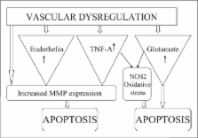
          Glaucoma, the second leading cause of blindness, is characterized by changes in the optic disc and visual field defects. The elevated intraocular pressure was considered the prime factor responsible for the glaucomatous optic neuropathy involving death of retinal ganglion cells and their axons. Extensive investigations into the pathophysiology of glaucoma now reveal the role of multiple factors in the development of retinal ganglion cell death. A better understanding of the pathophysiological mechanisms involved in the onset and progression of glaucomatous optic neuropathy is crucial in the development of better therapeutic options. This review is an effort to summarize the current concepts in the pathophysiology of glaucoma so that newer therapeutic targets can be recognized.

          The literature available in the National Medical Library and online Pubmed search engine was used for literature review.

          Related collections

          Most cited references127

          • Record: found
          • Abstract: found
          • Article: not found

          Age-related nuclear cataract-oxidation is the key.

          Age is by far the biggest risk factor for cataract, and it is sometimes assumed that cataract is simply an amplification of this aging process. This appears not to be the case, since the lens changes associated with aging and cataract are distinct. Oxidation is the hallmark of age-related nuclear (ARN) cataract. Loss of protein sulfhydryl groups, and the oxidation of methionine residues, are progressive and increase as the cataract worsens until >90% of cysteine and half the methionine residues are oxidised in the most advanced form. By contrast, there may be no significant oxidation of proteins in the centre of the lens with advancing age, even past age 80. The key factor in preventing oxidation seems to be the concentration of nuclear glutathione (GSH). Provided that nuclear GSH levels can be maintained above 2 mm, it appears that significant protein oxidation and posttranslational modification by reactive small molecules, such as ascorbate or UV filter degradation products, is not observed. Adequate coupling of the metabolically-active cortex, the source of antioxidants such as GSH, to the quiescent nucleus, is crucial especially since it would appear that the cortex remains viable in old lenses, and even possibly in ARN cataract lenses. Therefore it is vital to understand the reason for the onset of the lens barrier. This barrier, which becomes apparent in middle age, acts to impede the flow of small molecules between the cortex and the nucleus. The barrier, rather than nuclear compaction (which is not observed in human lenses), may contribute to the lowered concentration of GSH in the lens nucleus after middle age. By extending the residence time within the lens centre, the barrier also facilitates the decomposition of intrinsically unstable metabolites and may exacerbate the formation of H(2)O(2) in the nucleus. This hypothesis, which is based on the generation of reactive oxygen species and reactive molecules within the nucleus itself, shifts the focus away from theories for cataract that postulated a primary role for oxidants generated outside of the lens. Unfortunately, due to marked variability in the lenses of different species, there appears at present to be no ideal animal model system for studying human ARN cataract.
            Bookmark
            • Record: found
            • Abstract: found
            • Article: not found

            Retinal ganglion cell atrophy correlated with automated perimetry in human eyes with glaucoma.

            We measured the number and size of retinal ganglion cells from six human eyes with glaucoma. In each, the histologic findings were correlated with visual field results. Five age-matched normal eyes were studied for comparison. In general, there were fewer remaining large ganglion cells in retinal areas with atrophy. In the perifoveal area, however, no consistent pattern of cell loss by size was found. Our estimates suggest that visual field sensitivity in automated testing begins to decline soon after the initial loss of ganglion cells. Throughout the central 30 degrees of the retina, 20% of the normal number of cells were gone in locations with a 5-dB sensitivity loss, and 40% cell loss corresponded to a 10-dB decrease. There were some remaining ganglion cells in areas that had 0-dB sensitivity in the field test.
              Bookmark
              • Record: found
              • Abstract: found
              • Article: not found

              Vascular risk factors for primary open angle glaucoma: the Egna-Neumarkt Study.

              L Bonomi (2000)
              To assess the impact of vascular risk factors on the prevalence of primary open angle glaucoma. Population-based cross-sectional study. Four thousand two hundred ninety-seven patients more than 40 years of age underwent a complete ocular examination in the context of the Egna-Neumarkt Glaucoma Study. Ocular examinations were performed by trained, quality-controlled ophthalmologists according to a predefined standardized protocol including medical interview, blood pressure reading, applanation tonometry, computerized perimetry, and optic nerve head examination. Prevalences of ocular hypertension, primary open-angle glaucoma, normal-tension glaucoma, and other types of glaucoma were determined. Correlation coefficients were calculated for the association between systemic blood pressure and age-adjusted intraocular pressure (IOP) and between age and both intraocular and systemic blood pressures. Odds ratios were computed to assess the risk of primary open-angle glaucoma and normal-tension glaucoma in relation to systemic hypertension or antihypertensive medication, blood pressure levels, diastolic perfusion pressure, and a number of other cardiovascular risk factors. A positive correlation was found between systemic blood pressure and IOP, and an association was found between diagnosis of primary open-angle glaucoma and systemic hypertension. Lower diastolic perfusion pressure is associated with a marked, progressive increase in the frequency of hypertensive glaucoma. No relationship was found between systemic diseases of vascular origin and glaucoma. Our data are in line with those reported in other recent epidemiologic studies and show that reduced diastolic perfusion pressure is an important risk factor for primary open-angle glaucoma.
                Bookmark

                Author and article information

                Journal
                Indian J Ophthalmol
                IJO
                Indian Journal of Ophthalmology
                Medknow Publications (India )
                0301-4738
                1998-3689
                Jul-Aug 2009
                : 57
                : 4
                : 257-266
                Affiliations
                Department of Ocular Pharmacology, Delhi Institute of Pharmaceutical Sciences and Research, New Delhi, India
                [1 ]Dr. Rajendra Prasad Center for Ophthalmic Sciences, New Delhi, India
                Author notes
                Correspondence to Prof. SK Gupta, Department of Ocular Pharmacology, Delhi Institute of Pharmaceutical Sciences and Research, Pushp Vihar, Sector 3, MB Road, New Delhi, India. E-mail: skgup@ 123456hotmail.com
                Article
                IJO-57-257
                10.4103/0301-4738.53049
                2712693
                19574692
                53fd98f4-7e6b-4b8d-99db-f98a642fc700
                © Indian Journal of Ophthalmology

                This is an open-access article distributed under the terms of the Creative Commons Attribution License, which permits unrestricted use, distribution, and reproduction in any medium, provided the original work is properly cited.

                History
                : 19 December 2007
                : 26 May 2008
                Categories
                Review Article

                Ophthalmology & Optometry
                retinal ganglion cells,glaucoma,pathophysiology
                Ophthalmology & Optometry
                retinal ganglion cells, glaucoma, pathophysiology

                Comments

                Comment on this article