2
views
0
recommends
+1 Recommend
0 collections
    0
    shares
      • Record: found
      • Abstract: found
      • Article: found
      Is Open Access

      Translation elongation factor 2 depletion by siRNA in mouse liver leads to mTOR-independent translational upregulation of ribosomal protein genes

      research-article

      Read this article at

      Bookmark
          There is no author summary for this article yet. Authors can add summaries to their articles on ScienceOpen to make them more accessible to a non-specialist audience.

          Abstract

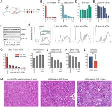
          Due to breakthroughs in RNAi and genome editing methods in the past decade, it is now easier than ever to study fine details of protein synthesis in animal models. However, most of our understanding of translation comes from unicellular organisms and cultured mammalian cells. In this study, we demonstrate the feasibility of perturbing protein synthesis in a mouse liver by targeting translation elongation factor 2 (eEF2) with RNAi. We were able to achieve over 90% knockdown efficacy and maintain it for 2 weeks effectively slowing down the rate of translation elongation. As the total protein yield declined, both proteomics and ribosome profiling assays showed robust translational upregulation of ribosomal proteins relative to other proteins. Although all these genes bear the TOP regulatory motif, the branch of the mTOR pathway responsible for translation regulation was not activated. Paradoxically, coordinated translational upregulation of ribosomal proteins only occurred in the liver but not in murine cell culture. Thus, the upregulation of ribosomal transcripts likely occurred via passive mTOR-independent mechanisms. Impaired elongation sequesters ribosomes on mRNA and creates a shortage of free ribosomes. This leads to preferential translation of transcripts with high initiation rates such as ribosomal proteins. Furthermore, severe eEF2 shortage reduces the negative impact of positively charged amino acids frequent in ribosomal proteins on ribosome progression.

          Related collections

          Most cited references30

          • Record: found
          • Abstract: found
          • Article: not found

          Regulation and function of ribosomal protein S6 kinase (S6K) within mTOR signalling networks.

          The ribosomal protein S6K (S6 kinase) represents an extensively studied effector of the TORC1 [TOR (target of rapamycin) complex 1], which possesses important yet incompletely defined roles in cellular and organismal physiology. TORC1 functions as an environmental sensor by integrating signals derived from diverse environmental cues to promote anabolic and inhibit catabolic cellular functions. mTORC1 (mammalian TORC1) phosphorylates and activates S6K1 and S6K2, whose first identified substrate was rpS6 (ribosomal protein S6), a component of the 40S ribosome. Studies over the past decade have uncovered a number of additional S6K1 substrates, revealing multiple levels at which the mTORC1-S6K1 axis regulates cell physiology. The results thus far indicate that the mTORC1-S6K1 axis controls fundamental cellular processes, including transcription, translation, protein and lipid synthesis, cell growth/size and cell metabolism. In the present review we summarize the regulation of S6Ks, their cellular substrates and functions, and their integration within rapidly expanding mTOR (mammalian TOR) signalling networks. Although our understanding of the role of mTORC1-S6K1 signalling in physiology remains in its infancy, evidence indicates that this signalling axis controls, at least in part, glucose homoeostasis, insulin sensitivity, adipocyte metabolism, body mass and energy balance, tissue and organ size, learning, memory and aging. As dysregulation of this signalling axis contributes to diverse disease states, improved understanding of S6K regulation and function within mTOR signalling networks may enable the development of novel therapeutics.
            Bookmark
            • Record: found
            • Abstract: found
            • Article: found
            Is Open Access

            Differential regulation of mTORC1 by leucine and glutamine

            The mechanistic target of rapamycin (mTOR) complex 1 (mTORC1) integrates environmental and intracellular signals to regulate cell growth. Amino acids stimulate mTORC1 activation at the lysosome in a manner thought to be dependent on the Rag small guanosine triphosphatases (GTPases), the Ragulator complex, and the vacuolar H+-adenosine triphosphatase (v-ATPase). We report that leucine and glutamine stimulate mTORC1 by Rag GTPase-dependent and -independent mechanisms, respectively. Glutamine promoted mTORC1 translocation to the lysosome in RagA and RagB knockout cells and required the v-ATPase but not the Ragulator. Furthermore, we identified the adenosine diphosphate ribosylation factor-1 GTPase to be required for mTORC1 activation and lysosomal localization by glutamine. Our results uncover a signaling cascade to mTORC1 activation independent of the Rag GTPases and suggest that mTORC1 is differentially regulated by specific amino acids.
              Bookmark
              • Record: found
              • Abstract: found
              • Article: not found

              A hierarchy of ATP-consuming processes in mammalian cells.

              The rates of different ATP-consuming reactions were measured in concanavalin A-stimulated thymocytes, a model system in which more than 80% of the ATP consumption can be accounted for. There was a clear hierarchy of the responses of different energy-consuming reactions to changes in energy supply: pathways of macromolecule biosynthesis (protein synthesis and RNA/DNA synthesis) were most sensitive to energy supply, followed by sodium cycling and then calcium cycling across the plasma membrane. Mitochondrial proton leak was the least sensitive to energy supply. Control analysis was used to quantify the relative control over ATP production exerted by the individual groups of ATP-consuming reactions. Control was widely shared; no block of reactions had more than one-third of the control. A fuller control analysis showed that there appeared to be a hierarchy of control over the flux through ATP: protein synthesis > RNA/DNA synthesis and substrate oxidation > Na+ cycling and Ca2+ cycling > other ATP consumers and mitochondrial proton leak. Control analysis also indicated that there was significant control over the rates of individual ATP consumers by energy supply. Each ATP consumer had strong control over its own rate but very little control over the rates of the other ATP consumers.
                Bookmark

                Author and article information

                Contributors
                mgerashchenko@bwh.harvard.edu
                kotelianskiv@gmail.com
                Journal
                Sci Rep
                Sci Rep
                Scientific Reports
                Nature Publishing Group UK (London )
                2045-2322
                23 September 2020
                23 September 2020
                2020
                : 10
                : 15473
                Affiliations
                [1 ]Division of Genetics, Department of Medicine, Brigham and Women’s Hospital, Harvard Medical School, Boston, MA 02115 USA
                [2 ]GRID grid.454320.4, ISNI 0000 0004 0555 3608, Skolkovo Institute of Science and Technology, ; Skolkovo, Moscow Region Russia
                [3 ]GRID grid.116068.8, ISNI 0000 0001 2341 2786, David H. Koch Institute for Integrative Cancer Research, , Massachusetts Institute of Technology, ; Cambridge, MA 02142 USA
                [4 ]GRID grid.38142.3c, ISNI 000000041936754X, Department of Cell Biology, , Harvard Medical School, ; Boston, MA 02115 USA
                [5 ]GRID grid.14476.30, ISNI 0000 0001 2342 9668, Belozersky Institute of Physico-Chemical Biology, Moscow State University, ; Moscow, 119992 Russia
                [6 ]GRID grid.14476.30, ISNI 0000 0001 2342 9668, Department of Chemistry, , Lomonosov Moscow State University, ; Moscow, Russia
                Article
                72399
                10.1038/s41598-020-72399-4
                7511953
                32968084
                55c15aec-8dab-4325-8beb-abe65213e9b6
                © The Author(s) 2020

                Open Access This article is licensed under a Creative Commons Attribution 4.0 International License, which permits use, sharing, adaptation, distribution and reproduction in any medium or format, as long as you give appropriate credit to the original author(s) and the source, provide a link to the Creative Commons licence, and indicate if changes were made. The images or other third party material in this article are included in the article's Creative Commons licence, unless indicated otherwise in a credit line to the material. If material is not included in the article's Creative Commons licence and your intended use is not permitted by statutory regulation or exceeds the permitted use, you will need to obtain permission directly from the copyright holder. To view a copy of this licence, visit http://creativecommons.org/licenses/by/4.0/.

                History
                : 26 March 2020
                : 15 July 2020
                Categories
                Article
                Custom metadata
                © The Author(s) 2020

                Uncategorized
                translation,rnai,inhibitory rna techniques,rnai therapy
                Uncategorized
                translation, rnai, inhibitory rna techniques, rnai therapy

                Comments

                Comment on this article