5
views
0
recommends
+1 Recommend
0 collections
    0
    shares
      • Record: found
      • Abstract: found
      • Article: found
      Is Open Access

      Increased interictal synchronicity of respiratory related brain pulsations in epilepsy

      research-article

      Read this article at

      Bookmark
          There is no author summary for this article yet. Authors can add summaries to their articles on ScienceOpen to make them more accessible to a non-specialist audience.

          Abstract

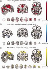
          Respiratory brain pulsations have recently been shown to drive electrophysiological brain activity in patients with epilepsy. Furthermore, functional neuroimaging indicates that respiratory brain pulsations have increased variability and amplitude in patients with epilepsy compared to healthy individuals. To determine whether the respiratory drive is altered in epilepsy, we compared respiratory brain pulsation synchronicity between healthy controls and patients. Whole brain fast functional magnetic resonance imaging was performed on 40 medicated patients with focal epilepsy, 20 drug-naïve patients and 102 healthy controls. Cerebrospinal fluid associated respiratory pulsations were used to generate individual whole brain respiratory synchronization maps, which were compared between groups. Finally, we analyzed the seizure frequency effect and diagnostic accuracy of the respiratory synchronization defect in epilepsy. Respiratory brain pulsations related to the verified fourth ventricle pulsations were significantly more synchronous in patients in frontal, periventricular and mid-temporal regions, while the seizure frequency correlated positively with synchronicity. The respiratory brain synchronicity had a good diagnostic accuracy (ROC AUC = 0.75) in discriminating controls from medicated patients. The elevated respiratory brain synchronicity in focal epilepsy suggests altered physiological effect of cerebrospinal fluid pulsations possibly linked to regional brain water dynamics involved with interictal brain physiology.

          Related collections

          Most cited references55

          • Record: found
          • Abstract: found
          • Article: not found

          A paravascular pathway facilitates CSF flow through the brain parenchyma and the clearance of interstitial solutes, including amyloid β.

          Because it lacks a lymphatic circulation, the brain must clear extracellular proteins by an alternative mechanism. The cerebrospinal fluid (CSF) functions as a sink for brain extracellular solutes, but it is not clear how solutes from the brain interstitium move from the parenchyma to the CSF. We demonstrate that a substantial portion of subarachnoid CSF cycles through the brain interstitial space. On the basis of in vivo two-photon imaging of small fluorescent tracers, we showed that CSF enters the parenchyma along paravascular spaces that surround penetrating arteries and that brain interstitial fluid is cleared along paravenous drainage pathways. Animals lacking the water channel aquaporin-4 (AQP4) in astrocytes exhibit slowed CSF influx through this system and a ~70% reduction in interstitial solute clearance, suggesting that the bulk fluid flow between these anatomical influx and efflux routes is supported by astrocytic water transport. Fluorescent-tagged amyloid β, a peptide thought to be pathogenic in Alzheimer's disease, was transported along this route, and deletion of the Aqp4 gene suppressed the clearance of soluble amyloid β, suggesting that this pathway may remove amyloid β from the central nervous system. Clearance through paravenous flow may also regulate extracellular levels of proteins involved with neurodegenerative conditions, its impairment perhaps contributing to the mis-accumulation of soluble proteins.
            Bookmark
            • Record: found
            • Abstract: found
            • Article: not found

            ILAE classification of the epilepsies: Position paper of the ILAE Commission for Classification and Terminology

            The International League Against Epilepsy (ILAE) Classification of the Epilepsies has been updated to reflect our gain in understanding of the epilepsies and their underlying mechanisms following the major scientific advances that have taken place since the last ratified classification in 1989. As a critical tool for the practicing clinician, epilepsy classification must be relevant and dynamic to changes in thinking, yet robust and translatable to all areas of the globe. Its primary purpose is for diagnosis of patients, but it is also critical for epilepsy research, development of antiepileptic therapies, and communication around the world. The new classification originates from a draft document submitted for public comments in 2013, which was revised to incorporate extensive feedback from the international epilepsy community over several rounds of consultation. It presents three levels, starting with seizure type, where it assumes that the patient is having epileptic seizures as defined by the new 2017 ILAE Seizure Classification. After diagnosis of the seizure type, the next step is diagnosis of epilepsy type, including focal epilepsy, generalized epilepsy, combined generalized, and focal epilepsy, and also an unknown epilepsy group. The third level is that of epilepsy syndrome, where a specific syndromic diagnosis can be made. The new classification incorporates etiology along each stage, emphasizing the need to consider etiology at each step of diagnosis, as it often carries significant treatment implications. Etiology is broken into six subgroups, selected because of their potential therapeutic consequences. New terminology is introduced such as developmental and epileptic encephalopathy. The term benign is replaced by the terms self-limited and pharmacoresponsive, to be used where appropriate. It is hoped that this new framework will assist in improving epilepsy care and research in the 21st century.
              Bookmark
              • Record: found
              • Abstract: found
              • Article: not found

              Threshold-free cluster enhancement: addressing problems of smoothing, threshold dependence and localisation in cluster inference.

              Many image enhancement and thresholding techniques make use of spatial neighbourhood information to boost belief in extended areas of signal. The most common such approach in neuroimaging is cluster-based thresholding, which is often more sensitive than voxel-wise thresholding. However, a limitation is the need to define the initial cluster-forming threshold. This threshold is arbitrary, and yet its exact choice can have a large impact on the results, particularly at the lower (e.g., t, z < 4) cluster-forming thresholds frequently used. Furthermore, the amount of spatial pre-smoothing is also arbitrary (given that the expected signal extent is very rarely known in advance of the analysis). In the light of such problems, we propose a new method which attempts to keep the sensitivity benefits of cluster-based thresholding (and indeed the general concept of "clusters" of signal), while avoiding (or at least minimising) these problems. The method takes a raw statistic image and produces an output image in which the voxel-wise values represent the amount of cluster-like local spatial support. The method is thus referred to as "threshold-free cluster enhancement" (TFCE). We present the TFCE approach and discuss in detail ROC-based optimisation and comparisons with cluster-based and voxel-based thresholding. We find that TFCE gives generally better sensitivity than other methods over a wide range of test signal shapes and SNR values. We also show an example on a real imaging dataset, suggesting that TFCE does indeed provide not just improved sensitivity, but richer and more interpretable output than cluster-based thresholding.
                Bookmark

                Author and article information

                Journal
                J Cereb Blood Flow Metab
                J Cereb Blood Flow Metab
                JCB
                spjcb
                Journal of Cerebral Blood Flow & Metabolism
                SAGE Publications (Sage UK: London, England )
                0271-678X
                1559-7016
                14 May 2022
                October 2022
                14 May 2022
                : 42
                : 10
                : 1840-1853
                Affiliations
                [1 ]Oulu Functional NeuroImaging (OFNI), Department of Diagnostic Radiology, Oulu University Hospital, Oulu, Finland
                [2 ]Medical Imaging, Physics and Technology (MIPT), Faculty of Medicine, University of Oulu, Oulu, Finland
                [3 ]Medical Research Center (MRC), Oulu, Finland
                [4 ]Center for Translational Neuromedicine, Department of Neurosurgery, University of Rochester Medical Center, Rochester, NY, USA
                [5 ]Center for Translational Neuromedicine, Faculty of Health and Medical Sciences, University of Copenhagen, Copenhagen, Denmark
                [6 ]Research Unit of Neuroscience, Neurology, University of Oulu, Oulu, Finland
                [7 ]Department of Neurology, Oulu University Hospital, Oulu, Finland
                [8 ]Department of Pediatric Neurology and Muscular Disease, University Medical Center Freiburg, Faculty of Medicine, University of Freiburg, Freiburg, Germany
                [9 ]Department of Paediatrics, Cumming School of Medicine, University of Calgary, Calgary, AB, Canada
                [10 ]Department of Neuroscience, Cumming School of Medicine, University of Calgary, Calgary, Canada
                [11 ]Hotchkiss Brain Institute and Alberta Children’s Hospital Research Institute, University of Calgary, Calgary, Canada
                [12 ]Department of Radiology, Medical Physics, University Medical Center Freiburg, Faculty of Medicine, University of Freiburg, Freiburg, Germany
                [13 ]Department of Radiology, Cumming School of Medicine, University of Calgary, Calgary, AB, Canada
                Author notes
                [*]Vesa Kiviniemi, Oulu Functional NeuroImaging (OFNI), Department of Diagnostic Radiology, Oulu University Hospital, P.O. Box 50, Oulu 90029, Finland. Email: vesa.kiviniemi@ 123456oulu.fi
                [*]Janne Kananen, Oulu Functional NeuroImaging (OFNI), Department of Diagnostic Radiology, Oulu University Hospital, P.O. Box 50, Oulu 90029, Finland. Email: janne.kananen@ 123456oulu.fi
                Author information
                https://orcid.org/0000-0001-6831-8056
                https://orcid.org/0000-0001-9403-4583
                https://orcid.org/0000-0001-5522-8334
                https://orcid.org/0000-0003-2884-6510
                Article
                10.1177_0271678X221099703
                10.1177/0271678X221099703
                9536129
                35570730
                562b419a-4975-4478-aa7e-707881b31471
                © The Author(s) 2022

                This article is distributed under the terms of the Creative Commons Attribution-NonCommercial 4.0 License ( https://creativecommons.org/licenses/by-nc/4.0/) which permits non-commercial use, reproduction and distribution of the work without further permission provided the original work is attributed as specified on the SAGE and Open Access pages ( https://us.sagepub.com/en-us/nam/open-access-at-sage).

                History
                : 26 July 2021
                : 2 March 2022
                : 7 April 2022
                Categories
                Original Articles
                Custom metadata
                ts2

                Neurosciences
                brain physiology,brain pulsations,epilepsy,fast fmri,respiratory synchronization
                Neurosciences
                brain physiology, brain pulsations, epilepsy, fast fmri, respiratory synchronization

                Comments

                Comment on this article