1
views
0
recommends
+1 Recommend
0 collections
    0
    shares
      • Record: found
      • Abstract: found
      • Article: found
      Is Open Access

      Retinal Cone Mosaic in sws1-Mutant Medaka ( Oryzias latipes), A Teleost

      research-article

      Read this article at

      Bookmark
          There is no author summary for this article yet. Authors can add summaries to their articles on ScienceOpen to make them more accessible to a non-specialist audience.

          Abstract

          Purpose

          Ablation of short single cones (SSCs) expressing short-wavelength-sensitive opsin (SWS1) is well analyzed in the field of regenerative retinal cells. In contrast with ablation studies, the phenomena caused by the complete deletion of SWS1 are less well-understood. To assess the effects of SWS1 deficiency on retinal structure, we established and analyzed sws1-mutant medaka.

          Methods

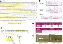
          To visualize SWS1, a monoclonal anti-SWS1 antibody and transgenic reporter fish ( Tg( sws1:mem-egfp)) were generated. We also developed a CRISPR/Cas-driven sws1-mutant line. Retinal structure of sws1 mutant was visualized using anti-SWS1, 1D4, and ZPR1 antibodies and coumarin derivatives and compared with wild type, Tg( sws1:mem-egfp) , and another opsin ( lws) mutant.

          Results

          Our rat monoclonal antibody specifically recognized medaka SWS1. S ws1 mutant retained regularly arranged cone mosaic as lws mutant and its SSCs had neither SWS1 nor long wavelength sensitive opsin. Depletion of sws1 did not affect the expression of long wavelength sensitive opsin, and vice versa. ZPR1 antibody recognized arrestin spread throughout double cones and long single cones in wild-type, transgenic, and sws1-mutant lines.

          Conclusions

          Comparative observation of sws1-mutant and wild-type retinas revealed that ZPR1 negativity is not a marker for SSCs with SWS1, but SSCs themselves. Loss of functional sws1 did not cause retinal degeneration, indicating that sws1 is not essential for cone mosaic development in medaka. Our two fish lines, one with visualized SWS1 and the other lacking functional SWS1, offer an opportunity to study neural network synapsing with SSCs and to clarify the role of SWS1 in vision.

          Related collections

          Most cited references88

          • Record: found
          • Abstract: found
          • Article: not found

          Fiji: an open-source platform for biological-image analysis.

          Fiji is a distribution of the popular open-source software ImageJ focused on biological-image analysis. Fiji uses modern software engineering practices to combine powerful software libraries with a broad range of scripting languages to enable rapid prototyping of image-processing algorithms. Fiji facilitates the transformation of new algorithms into ImageJ plugins that can be shared with end users through an integrated update system. We propose Fiji as a platform for productive collaboration between computer science and biology research communities.
            Bookmark
            • Record: found
            • Abstract: found
            • Article: found
            Is Open Access

            The EMBL-EBI search and sequence analysis tools APIs in 2019

            Abstract The EMBL-EBI provides free access to popular bioinformatics sequence analysis applications as well as to a full-featured text search engine with powerful cross-referencing and data retrieval capabilities. Access to these services is provided via user-friendly web interfaces and via established RESTful and SOAP Web Services APIs (https://www.ebi.ac.uk/seqdb/confluence/display/JDSAT/EMBL-EBI+Web+Services+APIs+-+Data+Retrieval). Both systems have been developed with the same core principles that allow them to integrate an ever-increasing volume of biological data, making them an integral part of many popular data resources provided at the EMBL-EBI. Here, we describe the latest improvements made to the frameworks which enhance the interconnectivity between public EMBL-EBI resources and ultimately enhance biological data discoverability, accessibility, interoperability and reusability.
              Bookmark
              • Record: found
              • Abstract: found
              • Article: not found

              Predicting transmembrane protein topology with a hidden Markov model: application to complete genomes.

              We describe and validate a new membrane protein topology prediction method, TMHMM, based on a hidden Markov model. We present a detailed analysis of TMHMM's performance, and show that it correctly predicts 97-98 % of the transmembrane helices. Additionally, TMHMM can discriminate between soluble and membrane proteins with both specificity and sensitivity better than 99 %, although the accuracy drops when signal peptides are present. This high degree of accuracy allowed us to predict reliably integral membrane proteins in a large collection of genomes. Based on these predictions, we estimate that 20-30 % of all genes in most genomes encode membrane proteins, which is in agreement with previous estimates. We further discovered that proteins with N(in)-C(in) topologies are strongly preferred in all examined organisms, except Caenorhabditis elegans, where the large number of 7TM receptors increases the counts for N(out)-C(in) topologies. We discuss the possible relevance of this finding for our understanding of membrane protein assembly mechanisms. A TMHMM prediction service is available at http://www.cbs.dtu.dk/services/TMHMM/. Copyright 2001 Academic Press.
                Bookmark

                Author and article information

                Journal
                Invest Ophthalmol Vis Sci
                Invest Ophthalmol Vis Sci
                IOVS
                Investigative Ophthalmology & Visual Science
                The Association for Research in Vision and Ophthalmology
                0146-0404
                1552-5783
                27 October 2022
                October 2022
                : 63
                : 11
                : 21
                Affiliations
                [1 ]Department of Chemical and Biological Sciences, Japan Women's University, Bunkyo-ku, Tokyo, Japan
                [2 ]Division of Molecular Genetics, Shigei Medical Research Institute, 2117 Yamada, Minami-ku, Okayama, Japan
                [3 ]Laboratory of Physiology, Atmosphere and Ocean Research Institute, The University of Tokyo, Kashiwa, Chiba, Japan
                [4 ]Laboratory of Bioresources/NIBB Center of the Interuniversity Bio-Backup Project, National Institute for Basic Biology, Okazaki, Aichi, Japan
                [5 ]Institute for Integrative Neurobiology and Department of Biology, Graduate School of Natural Science, Konan University, Kobe, Hyogo, Japan
                Author notes
                [* ]Correspondence: Megumi Matsuo and Shoji Fukamachi, Department of Chemical and Biological Sciences, Japan Women's University, Mejirodai 2-8-1, Bunkyo-ku, Tokyo 112-8681, Japan; mmatsuo@ 123456fc.jwu.ac.jp and fukamachi@ 123456fc.jwu.ac.jp .
                Article
                IOVS-22-35041
                10.1167/iovs.63.11.21
                9624274
                36301532
                5769d720-c9b6-4918-93db-2d8d762162d4
                Copyright 2022 The Authors

                This work is licensed under a Creative Commons Attribution 4.0 International License.

                History
                : 02 October 2022
                : 31 March 2022
                Page count
                Pages: 13
                Categories
                Retinal Cell Biology
                Retinal Cell Biology

                sws1 (short wavelength sensitive opsin),lws (long wavelength sensitive opsin),arrestin,cone photoreceptor,retinal degeneration

                Comments

                Comment on this article