44
views
0
recommends
+1 Recommend
0 collections
    0
    shares
      • Record: found
      • Abstract: found
      • Article: found
      Is Open Access

      Dexamethasone intravitreal implant in the treatment of diabetic macular edema

      review-article

      Read this article at

      Bookmark
          There is no author summary for this article yet. Authors can add summaries to their articles on ScienceOpen to make them more accessible to a non-specialist audience.

          Abstract

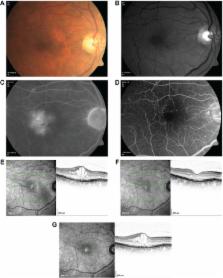
          Diabetic macular edema (DME) resembles a chronic, low-grade inflammatory reaction, and is characterized by blood–retinal barrier (BRB) breakdown and retinal capillary leakage. Corticosteroids are of therapeutic benefit because of their anti-inflammatory, antiangiogenic, and BRB-stabilizing properties. Delivery modes include periocular and intravitreal (via pars plana) injection. To offset the short intravitreal half-life of corticosteroid solutions (~3 hours) and the need for frequent intravitreal injections, sustained-release intravitreal corticosteroid implants have been developed. Dexamethasone intravitreal implant provides retinal drug delivery for ≤6 months and recently has been approved for use in the treatment of DME. Pooled findings (n=1,048) from two large-scale, randomized Phase III trials indicated that dexamethasone intravitreal implant (0.35 mg and 0.7 mg) administered at ≥6-month intervals produced sustained improvements in best-corrected visual acuity (BCVA) and macular edema. Significantly more patients showed a ≥15-letter gain in BCVA at 3 years with dexamethasone intravitreal implant 0.35 mg and 0.7 mg than with sham injection (18.4% and 22.2% vs 12.0%). Anatomical assessments showed rapid and sustained reductions in macular edema and slowing of retinopathy progression. Phase II study findings suggest that dexamethasone intravitreal implant is effective in focal, cystoid, and diffuse DME, in vitrectomized eyes, and in combination with laser therapy. Ocular complications of dexamethasone intravitreal implant in Phase III trials included cataract-related events (66.0% in phakic patients), intraocular pressure elevation ≥25 mmHg (29.7%), conjunctival hemorrhage (23.5%), vitreous hemorrhage (10.0%), macular fibrosis (8.3%), conjunctival hyperemia (7.2%), eye pain (6.1%), vitreous detachment (5.8%), and dry eye (5.8%); injection-related complications (eg, retinal tear/detachment, vitreous loss, endophthalmitis) were infrequent (<2%). Dexamethasone intravitreal implant offers a viable treatment option for DME, especially in cases that are persistent or treatment (anti-vascular endothelial growth factor/laser) refractory.

          Related collections

          Most cited references73

          • Record: found
          • Abstract: found
          • Article: not found

          A central role for inflammation in the pathogenesis of diabetic retinopathy.

          Diabetic retinopathy is a leading cause of adult vision loss and blindness. Much of the retinal damage that characterizes the disease results from retinal vascular leakage and nonperfusion. Diabetic retinal vascular leakage, capillary nonperfusion, and endothelial cell damage are temporary and spatially associated with retinal leukocyte stasis in early experimental diabetes. Retinal leukostasis increases within days of developing diabetes and correlates with the increased expression of retinal intercellular adhesion molecule-1 (ICAM-1) and CD18. Mice deficient in the genes encoding for the leukocyte adhesion molecules CD18 and ICAM-1 were studied in two models of diabetic retinopathy with respect to the long-term development of retinal vascular lesions. CD18-/- and ICAM-1-/- mice demonstrate significantly fewer adherent leukocytes in the retinal vasculature at 11 and 15 months after induction of diabetes with STZ. This condition is associated with fewer damaged endothelial cells and lesser vascular leakage. Galactosemia of up to 24 months causes pericyte and endothelial cell loss and formation of acellular capillaries. These changes are significantly reduced in CD18- and ICAM-1-deficient mice. Basement membrane thickening of the retinal vessels is increased in long-term galactosemic animals independent of the genetic strain. Here we show that chronic, low-grade subclinical inflammation is responsible for many of the signature vascular lesions of diabetic retinopathy. These data highlight the central and causal role of adherent leukocytes in the pathogenesis of diabetic retinopathy. They also underscore the potential utility of anti-inflammatory treatment in diabetic retinopathy.
            Bookmark
            • Record: found
            • Abstract: found
            • Article: not found

            Diabetic retinopathy: seeing beyond glucose-induced microvascular disease.

            Diabetic retinopathy remains a frightening prospect to patients and frustrates physicians. Destruction of damaged retina by photocoagulation remains the primary treatment nearly 50 years after its introduction. The diabetes pandemic requires new approaches to understand the pathophysiology and improve the detection, prevention, and treatment of retinopathy. This perspective considers how the unique anatomy and physiology of the retina may predispose it to the metabolic stresses of diabetes. The roles of neural retinal alterations and impaired retinal insulin action in the pathogenesis of early retinopathy and the mechanisms of vision loss are emphasized. Potential means to overcome limitations of current animal models and diagnostic testing are also presented with the goal of accelerating therapies to manage retinopathy in the face of ongoing diabetes.
              Bookmark
              • Record: found
              • Abstract: found
              • Article: not found

              Pharmacokinetics and pharmacodynamics of a sustained-release dexamethasone intravitreal implant.

              To determine the pharmacokinetics and pharmacodynamics of a sustained-release dexamethasone (DEX) intravitreal implant (Ozurdex; Allergan, Inc.). Thirty-four male monkeys (Macaca fascicularis) received bilateral 0.7-mg DEX implants. Blood, vitreous humor, and retina samples were collected at predetermined intervals up to 270 days after administration. DEX was quantified by liquid chromatography-tandem mass spectrometry, and cytochrome P450 3A8 (CYP3A8) gene expression was analyzed by real-time reverse transcription-polymerase chain reaction. DEX was detected in the retina and vitreous humor for 6 months, with peak concentrations during the first 2 months. After 6 months, DEX was below the limit of quantitation. The C(max) (T(max)) and AUC for the retina were 1110 ng/g (day 60) and 47,200 ng · d/g, and for the vitreous humor were 213 ng/mL (day 60) and 11,300 ng · d/mL, respectively. The C(max) (T(max)) of DEX in plasma was 1.11 ng/mL (day 60). Compared with the level in the control eyes (no DEX implant), CYP3A8 expression in the retina was upregulated threefold up to 6 months after injection of the implant (0.969 ± 0.0565 vs. 3.07 ± 0.438; P < 0.05 up to 2-month samples). The in vivo release profile of the DEX implant in an animal eye was similar to the pharmacokinetics achieved with pulse administration of corticosteroids (high initial drug concentration, followed by a prolonged period of low concentration). These results are consistent with those in clinical studies supporting the use of the DEX implant for the extended management of posterior segment diseases.
                Bookmark

                Author and article information

                Journal
                Clin Ophthalmol
                Clin Ophthalmol
                Clinical Ophthalmology
                Clinical Ophthalmology (Auckland, N.Z.)
                Dove Medical Press
                1177-5467
                1177-5483
                2015
                16 July 2015
                : 9
                : 1321-1335
                Affiliations
                [1 ]Retinal Consultants of Arizona, Phoenix, AZ, Los Angeles, CA, USA
                [2 ]Department of Ophthalmology, Keck School of Medicine, University of Southern California, Los Angeles, CA, USA
                [3 ]Department of Ophthalmology, University Vita-Salute Scientific Institute San Raffaele, Milan, Italy
                [4 ]Department of Ophthalmology, Tel Aviv Medical Center and Sackler Faculty of Medicine, Tel Aviv University, Tel Aviv, Israel
                Author notes
                Correspondence: Pravin U Dugel, Retinal Consultants of Arizona, 1101 East Missouri Avenue, PO Box 32530, Phoenix, AZ 85014-2709, USA, Tel +1 602 222 2221, Fax +1 602 682 2740, Email pdugel@ 123456gmail.com
                Article
                opth-9-1321
                10.2147/OPTH.S79948
                4509543
                26213460
                58202332-b6e8-4081-a1f8-0c1994e5d5fe
                © 2015 Dugel et al. This work is published by Dove Medical Press Limited, and licensed under Creative Commons Attribution – Non Commercial (unported, v3.0) License

                The full terms of the License are available at http://creativecommons.org/licenses/by-nc/3.0/. Non-commercial uses of the work are permitted without any further permission from Dove Medical Press Limited, provided the work is properly attributed.

                History
                Categories
                Review

                Ophthalmology & Optometry
                corticosteroids,dexamethasone,intravitreal,implant,macular edema,diabetic retinopathy

                Comments

                Comment on this article