11
views
0
recommends
+1 Recommend
0 collections
    0
    shares
      • Record: found
      • Abstract: found
      • Article: found
      Is Open Access

      A functional connection between dyskerin and energy metabolism

      brief-report

      Read this article at

      Bookmark
          There is no author summary for this article yet. Authors can add summaries to their articles on ScienceOpen to make them more accessible to a non-specialist audience.

          Abstract

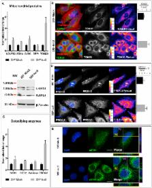
          The human DKC1 gene encodes dyskerin, an evolutionarily conserved nuclear protein whose overexpression represents a common trait of many types of aggressive sporadic cancers. As a crucial component of the nuclear H/ACA snoRNP complexes, dyskerin is involved in a variety of essential processes, including telomere maintenance, splicing efficiency, ribosome biogenesis, snoRNAs stabilization and stress response. Although multiple minor dyskerin splicing isoforms have been identified, their functions remain to be defined. Considering that low-abundance splice variants could contribute to the wide functional repertoire attributed to dyskerin, possibly having more specialized tasks or playing significant roles in changing cell status, we investigated in more detail the biological roles of a truncated dyskerin isoform that lacks the C-terminal nuclear localization signal and shows a prevalent cytoplasmic localization. Here we show that this dyskerin variant can boost energy metabolism and improve respiration, ultimately conferring a ROS adaptive response and a growth advantage to cells. These results reveal an unexpected involvement of DKC1 in energy metabolism, highlighting a previously underscored role in the regulation of metabolic cell homeostasis.

          Graphical abstract

          Highlights

          • Human dyskerin is an evolutionary conserved component of nuclear H/ACA snoRNPs.

          • The functional role of a truncated dyskerin isoform (Iso3) is analyzed.

          • Iso3 overexpression boosts energy metabolism and induces a ROS adaptive response.

          • Iso3 connects dyskerin with mitochondrial functionality and redox homeostasis.

          Related collections

          Most cited references50

          • Record: found
          • Abstract: found
          • Article: not found

          Pseudouridine profiling reveals regulated mRNA pseudouridylation in yeast and human cells

          Post-transcriptional modification of RNA nucleosides occurs in all living organisms. Pseudouridine, the most abundant modified nucleoside in non-coding RNAs 1 , enhances the function of transfer RNA and ribosomal RNA by stabilizing RNA structure 2–8 . mRNAs were not known to contain pseudouridine, but artificial pseudouridylation dramatically affects mRNA function – it changes the genetic code by facilitating non-canonical base pairing in the ribosome decoding center 9,10 . However, without evidence of naturally occurring mRNA pseudouridylation, its physiological was unclear. Here we present a comprehensive analysis of pseudouridylation in yeast and human RNAs using Pseudo-seq, a genome-wide, single-nucleotide-resolution method for pseudouridine identification. Pseudo-seq accurately identifies known modification sites as well as 100 novel sites in non-coding RNAs, and reveals hundreds of pseudouridylated sites in mRNAs. Genetic analysis allowed us to assign most of the new modification sites to one of seven conserved pseudouridine synthases, Pus1–4, 6, 7 and 9. Notably, the majority of pseudouridines in mRNA are regulated in response to environmental signals, such as nutrient deprivation in yeast and serum starvation in human cells. These results suggest a mechanism for the rapid and regulated rewiring of the genetic code through inducible mRNA modifications. Our findings reveal unanticipated roles for pseudouridylation and provide a resource for identifying the targets of pseudouridine synthases implicated in human disease 11–13 .
            Bookmark
            • Record: found
            • Abstract: found
            • Article: not found

            Mitochondrial complex I inhibitor rotenone induces apoptosis through enhancing mitochondrial reactive oxygen species production.

            Inhibition of mitochondrial respiratory chain complex I by rotenone had been found to induce cell death in a variety of cells. However, the mechanism is still elusive. Because reactive oxygen species (ROS) play an important role in apoptosis and inhibition of mitochondrial respiratory chain complex I by rotenone was thought to be able to elevate mitochondrial ROS production, we investigated the relationship between rotenone-induced apoptosis and mitochondrial reactive oxygen species. Rotenone was able to induce mitochondrial complex I substrate-supported mitochondrial ROS production both in isolated mitochondria from HL-60 cells as well as in cultured cells. Rotenone-induced apoptosis was confirmed by DNA fragmentation, cytochrome c release, and caspase 3 activity. A quantitative correlation between rotenone-induced apoptosis and rotenone-induced mitochondrial ROS production was identified. Rotenone-induced apoptosis was inhibited by treatment with antioxidants (glutathione, N-acetylcysteine, and vitamin C). The role of rotenone-induced mitochondrial ROS in apoptosis was also confirmed by the finding that HT1080 cells overexpressing magnesium superoxide dismutase were more resistant to rotenone-induced apoptosis than control cells. These results suggest that rotenone is able to induce apoptosis via enhancing the amount of mitochondrial reactive oxygen species production.
              Bookmark
              • Record: found
              • Abstract: found
              • Article: not found

              PGC1α and mitochondrial metabolism--emerging concepts and relevance in ageing and neurodegenerative disorders.

              PGC1α is a transcriptional coactivator that is a central inducer of mitochondrial biogenesis in cells. Recent work highlighted that PGC1α can also modulate the composition and functions of individual mitochondria. Therefore, it is emerging that PGC1α is controlling global oxidative metabolism by performing two types of remodelling: (1) cellular remodelling through mitochondrial biogenesis, and (2) organelle remodelling through alteration in the intrinsic properties of mitochondria. The elevated oxidative metabolism associated with increased PGC1α activity could be accompanied by an increase in reactive oxygen species (ROS) that are primarily generated by mitochondria. However, increasing evidence suggests that this is not the case, as PGC1α is also a powerful regulator of ROS removal by increasing the expression of numerous ROS-detoxifying enzymes. Therefore, PGC1α, by controlling both the induction of mitochondrial metabolism and the removal of its ROS by-products, would elevate oxidative metabolism and minimize the impact of ROS on cell physiology. In this Commentary, we discuss how the biogenesis and remodelling of mitochondria that are elicited by PGC1α contribute to an increase in oxidative metabolism and the preservation of ROS homeostasis. Finally, we examine the importance of these findings in ageing and neurodegenerative disorders, conditions that are associated with impaired mitochondrial functions and ROS balance.
                Bookmark

                Author and article information

                Contributors
                Journal
                Redox Biol
                Redox Biol
                Redox Biology
                Elsevier
                2213-2317
                06 November 2017
                April 2018
                06 November 2017
                : 14
                : 557-565
                Affiliations
                [a ]Department of Biology, University of Naples “Federico II”, Complesso Universitario Monte Santangelo, via Cinthia, 80126 Napoli, Italy
                [b ]Istituto Italiano di Tecnologia, IIT@CRIB, Largo Barsanti e Matteucci 53, 80125 Napoli, Italy
                [c ]Institute for Cell and Molecular Biosciences, Campus for Ageing and Vitality, University of Newcastle, Newcastle-upon-Tyne NE4 5PL, United Kingdom
                [d ]Centro di Ricerca Interdipartimentale sui Biomateriali CRIB, Università di Napoli Federico II, Piazzale Tecchio 80, 80125 Napoli, Italy
                Author notes
                [* ]Correspondence to: Department of Biology, University of Naples, via Cinthia, 80126 Napoli, Italy.Department of Biology, University of Naplesvia CinthiaNapoli80126Italy alberto.angrisani@ 123456unina.it mfuria@ 123456unina.it
                [1]

                Present address: Dipartimento Medico-Chirurgico di Internistica Clinica e Sperimentale "F. Magrassi e A. Lanzara. Laboratorio Oncologia Molecolare, Università della Campania Luigi Vanvitelli, Via S. Pansini 5, 80131, Napoli-Italy.

                [2]

                Present address: NICHD (National Institute of Child Health and Human Development)-Section on Metabolic Regulation-NIH-35 Convent DR, Bethesda, MD 20814, USA.

                Article
                S2213-2317(17)30547-5
                10.1016/j.redox.2017.11.003
                5684492
                29132127
                599537a0-5c3a-4d5c-b258-000763ae8989
                © 2017 The Authors

                This is an open access article under the CC BY-NC-ND license (http://creativecommons.org/licenses/by-nc-nd/4.0/).

                History
                : 26 July 2017
                : 12 October 2017
                : 3 November 2017
                Categories
                Short Communication

                dkc1,energy metabolism,mitochondria,ros signaling,prdx-2
                dkc1, energy metabolism, mitochondria, ros signaling, prdx-2

                Comments

                Comment on this article