50
views
0
recommends
+1 Recommend
0 collections
    0
    shares
      • Record: found
      • Abstract: found
      • Article: found
      Is Open Access

      Claudin-1, -2 and -3 Are Selectively Expressed in the Epithelia of the Choroid Plexus of the Mouse from Early Development and into Adulthood While Claudin-5 is Restricted to Endothelial Cells

      research-article

      Read this article at

      Bookmark
          There is no author summary for this article yet. Authors can add summaries to their articles on ScienceOpen to make them more accessible to a non-specialist audience.

          Abstract

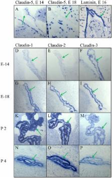
          A primary function of epithelial and endothelial monolayers is the formation of barriers that separate tissues into functional compartments. Tight junctions (TJs) seal the intercellular space between the single cells of a monolayer. TJs thus contribute importantly to the homeostasis of the cerebrospinal fluid as they help in maintaining the blood-brain barrier (BBB) and the blood-cerebrospinal fluid barrier (CSF). The composition of TJs differs by its localization as well as the stage of development according to its respective function. Claudin-3 is typically present in the epithelia and has been claimed to be a constituent of the BBB. It is, however, notoriously difficult to demonstrate its expression in endothelial cells of the brain vasculature at the morphological level by means of immunohistochemical techniques. Using an improved fixation strategy (4% paraformaldehyde at pH 11, in the presence of EDTA) and the sensitive alkaline phosphatase as a detection system, we show that claudin-3 is present in mouse epithelia from embryonic day 14 onwards. In brain, it is restricted to the anlage of choroid plexus in the ventricles, together with claudin-1 and -2. In adult mice, it is clearly delineating the epithelium of the choroid plexus in the lateral and fourth ventricles. In contrast, in cerebral blood vessels claudin-3 as well as claudin-1 and -2 are absent in cerebral blood vessels during all developmental stages up to adulthood. Rather, the BBB is characterized by the presence of claudin-5, ZO-1 and occludin. Thus, in mice claudin-3 is an important constituent of TJ in the embryonic and in the adult choroid plexus.

          Related collections

          Most cited references33

          • Record: found
          • Abstract: found
          • Article: not found

          Complex phenotype of mice lacking occludin, a component of tight junction strands.

          Occludin is an integral membrane protein with four transmembrane domains that is exclusively localized at tight junction (TJ) strands. Here, we describe the generation and analysis of mice carrying a null mutation in the occludin gene. Occludin -/- mice were born with no gross phenotype in the expected Mendelian ratios, but they showed significant postnatal growth retardation. Occludin -/- males produced no litters with wild-type females, whereas occludin -/- females produced litters normally when mated with wild-type males but did not suckle them. In occludin -/- mice, TJs themselves did not appear to be affected morphologically, and the barrier function of intestinal epithelium was normal as far as examined electrophysiologically. However, histological abnormalities were found in several tissues, i.e., chronic inflammation and hyperplasia of the gastric epithelium, calcification in the brain, testicular atrophy, loss of cytoplasmic granules in striated duct cells of the salivary gland, and thinning of the compact bone. These phenotypes suggested that the functions of TJs as well as occludin are more complex than previously supposed.
            Bookmark
            • Record: found
            • Abstract: found
            • Article: not found

            A Single Gene Product, Claudin-1 or -2, Reconstitutes Tight Junction Strands and Recruits Occludin in Fibroblasts

            Three integral membrane proteins, clau- din-1, -2, and occludin, are known to be components of tight junction (TJ) strands. To examine their ability to form TJ strands, their cDNAs were introduced into mouse L fibroblasts lacking TJs. Immunofluorescence microscopy revealed that both FLAG-tagged claudin-1 and -2 were highly concentrated at cell contact sites as planes through a homophilic interaction. In freeze-fracture replicas of these contact sites, well-developed networks of strands were identified that were similar to TJ strand networks in situ and were specifically labeled with anti-FLAG mAb. In glutaraldehyde-fixed samples, claudin-1–induced strands were largely associated with the protoplasmic (P) face as mostly continuous structures, whereas claudin-2–induced strands were discontinuous at the P face with complementary grooves at the extracellular (E) face which were occupied by chains of particles. Although occludin was also concentrated at cell contact sites as dots through its homophilic interaction, freeze-fracture replicas identified only a small number of short strands that were labeled with anti-occludin mAb. However, when occludin was cotransfected with claudin-1, it was concentrated at cell contact sites as planes to be incorporated into well- developed claudin-1–based strands. These findings suggested that claudin-1 and -2 are mainly responsible for TJ strand formation, and that occludin is an accessory protein in some function of TJ strands.
              Bookmark
              • Record: found
              • Abstract: found
              • Article: not found

              The blood-brain barrier and cancer: transporters, treatment, and Trojan horses.

              Despite scientific advances in understanding the causes and treatment of human malignancy, a persistent challenge facing basic and clinical investigators is how to adequately treat primary and metastatic brain tumors. The blood-brain barrier is a physiologic obstruction to the delivery of systemic chemotherapy to the brain parenchyma and central nervous system (CNS). A number of physiologic properties make the endothelium in the CNS distinct from the vasculature found in the periphery. Recent evidence has shown that a critical aspect of this barrier is composed of xenobiotic transporters which extrude substrates from the brain into the cerebrospinal fluid and systemic circulation. These transporters also extrude drugs and toxins if they gain entry into the cytoplasm of brain endothelial cells before they enter the brain. This review highlights the properties of the blood-brain barrier, including the location, function, and relative importance of the drug transporters that maintain this barrier. Primary and metastatic brain malignancy can compromise this barrier, allowing some access of chemotherapy treatment to reach the tumor. The responsiveness of brain tumors to systemic treatment found in past clinical research is discussed, as are possible explanations as to why CNS tumors are nonetheless able to evade therapy. Finally, strategies to overcome this barrier and better deliver chemotherapy into CNS tumors are presented.
                Bookmark

                Author and article information

                Contributors
                Journal
                Front Neuroanat
                Front Neuroanat
                Front. Neuroanat.
                Frontiers in Neuroanatomy
                Frontiers Media S.A.
                1662-5129
                22 February 2016
                2016
                : 10
                : 16
                Affiliations
                [1]Department of Biomedicine, Section of Functional Neuroanatomy, University of Basel Basel, Switzerland
                Author notes

                Edited by: Zoltan F. Kisvarday, University of Debrecen, Hungary

                Reviewed by: Nobuaki Tamamaki, Kumamoto University, Japan; Tom Reese, National Institutes of Health, USA; Patricia Gaspar, Institut national de la santé et de la recherche médicale, France

                *Correspondence: Cordula Nitsch cordula.nitsch@ 123456unibas.ch

                Present address: Sophorn Chip, Department of Neurology, University of California San Francisco, San Francisco, CA, USA

                Article
                10.3389/fnana.2016.00016
                4761916
                26941614
                5ac50c3d-861c-42bf-b452-48c3efe59e4a
                Copyright © 2016 Steinemann, Galm, Chip, Nitsch and Maly.

                This is an open-access article distributed under the terms of the Creative Commons Attribution License (CC BY). The use, distribution and reproduction in other forums is permitted, provided the original author(s) or licensor are credited and that the original publication in this journal is cited, in accordance with accepted academic practice. No use, distribution or reproduction is permitted which does not comply with these terms.

                History
                : 05 October 2015
                : 05 February 2016
                Page count
                Figures: 6, Tables: 0, Equations: 0, References: 47, Pages: 10, Words: 5988
                Categories
                Neuroscience
                Original Research

                Neurosciences
                blood-brain barrier,claudin-5,tight junctions,zonula occludens,occludin,ependyma,alkaline phosphatase

                Comments

                Comment on this article