18
views
0
recommends
+1 Recommend
0 collections
    0
    shares
      • Record: found
      • Abstract: found
      • Article: found
      Is Open Access

      Volumetric and Fatty Infiltration Imbalance of Deep Paravertebral Muscles in Adolescent Idiopathic Scoliosis

      research-article

      Read this article at

      Bookmark
          There is no author summary for this article yet. Authors can add summaries to their articles on ScienceOpen to make them more accessible to a non-specialist audience.

          Abstract

          Background

          Several studies have described the differences in electromyographic activity and histological changes of paravertebral muscles in patients with adolescent idiopathic scoliosis (AIS). However, there is little knowledge about the muscle volumetric and fatty infiltration imbalance of patients with AIS.

          Material/Methods

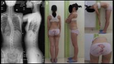
          Thirty-four patients with AIS were evaluated with standardized anteroposterior (AP) and lateral standing films for the location and direction of the apex of scoliosis, coronal Cobb angle, apex vertebra translation, and thoracic kyphosis; and with magnetic resonance imaging (MRI) scan of the spine at the level of T4–L1. The muscle volume and fatty infiltration rate of bilateral deep paravertebral muscles at the level of upper end, apex, and lower end vertebra were measured.

          Results

          All patients had major thoracic curve with apex of curves on the right side. The muscle volume on the convex side was larger relative to the concave side at the three levels, while the fatty infiltration rate was significantly higher on the concave side. The difference index of the muscle volume was significantly larger at the apex vertebra level than at the upper end vertebra level ( p=0.002) or lower end vertebra level ( p<0.001). The difference index of muscle volume correlated with apex vertebra translation (r=−0.749, p=0.032), and the difference index of fatty involution correlated with apex vertebra translation (r=0.727, p=0.041) and Cobb angle (r=0.866, p=0.005).

          Conclusions

          Our findings demonstrated significant imbalance of muscle volume and fatty infiltration in deep paravertebral muscles of AIS patients. Moreover, these changes affected different vertebra levels, with the most imbalance of muscle volume at the apex vertebra. We interpreted this as morphological changes corresponding with known altered muscle function of AIS.

          Related collections

          Most cited references30

          • Record: found
          • Abstract: found
          • Article: not found

          Effects of bracing in adolescents with idiopathic scoliosis.

          The role of bracing in patients with adolescent idiopathic scoliosis who are at risk for curve progression and eventual surgery is controversial. We conducted a multicenter study that included patients with typical indications for bracing due to their age, skeletal immaturity, and degree of scoliosis. Both a randomized cohort and a preference cohort were enrolled. Of 242 patients included in the analysis, 116 were randomly assigned to bracing or observation, and 126 chose between bracing and observation. Patients in the bracing group were instructed to wear the brace at least 18 hours per day. The primary outcomes were curve progression to 50 degrees or more (treatment failure) and skeletal maturity without this degree of curve progression (treatment success). The trial was stopped early owing to the efficacy of bracing. In an analysis that included both the randomized and preference cohorts, the rate of treatment success was 72% after bracing, as compared with 48% after observation (propensity-score-adjusted odds ratio for treatment success, 1.93; 95% confidence interval [CI], 1.08 to 3.46). In the intention-to-treat analysis, the rate of treatment success was 75% among patients randomly assigned to bracing, as compared with 42% among those randomly assigned to observation (odds ratio, 4.11; 95% CI, 1.85 to 9.16). There was a significant positive association between hours of brace wear and rate of treatment success (P<0.001). Bracing significantly decreased the progression of high-risk curves to the threshold for surgery in patients with adolescent idiopathic scoliosis. The benefit increased with longer hours of brace wear. (Funded by the National Institute of Arthritis and Musculoskeletal and Skin Diseases and others; BRAIST ClinicalTrials.gov number, NCT00448448.).
            Bookmark
            • Record: found
            • Abstract: found
            • Article: not found

            A genome-wide association study identifies common variants near LBX1 associated with adolescent idiopathic scoliosis.

            Adolescent idiopathic scoliosis is a pediatric spinal deformity affecting 2-3% of school-age children worldwide(1). Genetic factors have been implicated in its etiology(2). Through a genome-wide association study (GWAS) and replication study involving a total of 1,376 Japanese females with adolescent idiopathic scoliosis and 11,297 female controls, we identified a locus at chromosome 10q24.31 associated with adolescent idiopathic scoliosis susceptibility. The most significant SNP (rs11190870; combined P = 1.24 × 10(-19); odds ratio (OR) = 1.56) is located near LBX1 (encoding ladybird homeobox 1). The identification of this susceptibility locus provides new insights into the pathogenesis of adolescent idiopathic scoliosis.
              Bookmark
              • Record: found
              • Abstract: not found
              • Article: not found

              Clinical practice. Idiopathic scoliosis in adolescents.

              M Hresko (2013)
                Bookmark

                Author and article information

                Journal
                Med Sci Monit
                Med. Sci. Monit
                Medical Science Monitor
                Medical Science Monitor : International Medical Journal of Experimental and Clinical Research
                International Scientific Literature, Inc.
                1234-1010
                1643-3750
                2017
                02 May 2017
                : 23
                : 2089-2095
                Affiliations
                [1 ]Department of Orthopedics, Changzheng Hospital, Second Military Medical University, Shanghai, P.R. China
                [2 ]Department of Orthopedics, Shanghai General Hospital, Shanghai Jiao Tong University, Shanghai, P.R. China
                Author notes
                Corresponding Authors: Xuhui Zhou, e-mail: xhzhouspine@ 123456163.com or Rui Gao, e-mail: rgaospine@ 123456163.com
                [A]

                Study Design

                [B]

                Data Collection

                [C]

                Statistical Analysis

                [D]

                Data Interpretation

                [E]

                Manuscript Preparation

                [F]

                Literature Search

                [G]

                Funds Collection

                [*]

                Heng Jiang, Yichen Meng and Xinmeng Jin contributed equally to this work

                Article
                902455
                10.12659/MSM.902455
                5424650
                28461686
                5b77efd7-4a1a-4f03-a1eb-3fd7460aa78c
                © Med Sci Monit, 2017

                This work is licensed under Creative Common Attribution-NonCommercial-NoDerivatives 4.0 International ( CC BY-NC-ND 4.0)

                History
                : 20 November 2016
                : 12 December 2016
                Categories
                Clinical Research

                magnetic resonance imaging,muscular atrophy,paraspinal muscles,scoliosis

                Comments

                Comment on this article