- Record: found
- Abstract: found
- Article: found
Editorial for the Special Issue on MEMS Technology for Biomedical Imaging Applications
editorial
Read this article at
There is no author summary for this article yet. Authors can add summaries to their articles on ScienceOpen to make them more accessible to a non-specialist audience.
Abstract
Biomedical imaging is the key technique and process to create informative images of
the human body or other organic structures for clinical purposes or medical science.
Micro-electro-mechanical systems (MEMS) technology has demonstrated enormous potential
in biological imaging applications due to its outstanding advantages of, for instance,
miniaturization, high speed, higher resolution, and convenience of batch fabrication.
There are many advancements and breakthroughs developing in the academic community,
and there are a few challenges raised accordingly upon the designs, structures, fabrication,
integration, and applications of MEMS for all kinds of biomedical imaging. This Special
Issue of Micromachines, entitled “MEMS Technology for Biomedical Imaging Applications”,
contains 13 papers (nine articles and four reviews) highlighting recent advances in
the field of biomedical imaging and covering broad topics from the key components
to the applications of various imaging systems.
In the area of ultrasonic transducers, Brenner et al. reviewed the capacitive micromachined
transducers at all levels: Theory and modeling methods, fabrication technologies,
system integration, as well as imaging applications [1]. Future trends for capacitive
micromachined ultrasonic transducers and their impact within the broad field of biomedical
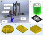
imaging were also discussed. Work by Chen et al. was aimed to provide a piezoelectric
array to improve the acoustic field and spatial resolution in medical ultrasonic imaging
[2]. Photocurable resin and nano ceramic particles can be 3D-printed into different
concentric elements to consist annular piezoelectric arrays, which are capable of
tuning the focus zone and lateral resolution. The design, fabrication, and characterization
of a tightly focused high frequency needle-type ultrasonic transducer made by Co-doped
Na0.5Bi4.5Ti4O15 ceramics was demonstrated by Fei et al. [3]. Li et al. also presented
tightly focused ultrasonic transducers, which were designed using aluminum nitride
thin film as piezoelectric element and using silicon lens for focusing [4]. In addition,
a custom designed integrated circuit combining a high frequency wideband low noise
amplifier with a common-source and common-gate structure was used to process the ultrasonic
medical echo signal with low noise figure, high gain, and good linearity.
This issue has two papers in the field of photoacoustic imaging. Lee et al. reviewed
cutting-edge MEMS technologies for photoacoustic imaging and summarizes the recent
advances of scanning mirrors and detectors [5]. Conventional silicon and water immersible
scanning mirrors were introduced respectively, followed by micromachined transducers,
microring resonators, as well as silicon acoustic delay lines and multiplexers. In
the work of Qi et al., an optical resolution photoacoustic microscopy system based
on a MEMS scanning mirror was proposed [6]. The mirror was used to achieve raster
scanning of the excitation optical focus and the photoacoustic signal was detected
by a flat transducer in the system.
Two papers on microendoscopy are included in this issue. Qiu et al. presented a review
of the advancements of MEMS actuators for optical microendoscopy, including optical
coherence tomography, optical resolution photoacoustic microscopy, confocal, multiphoton,
and fluorescence wide-field microendoscopy [7]. The work of Yang et al. provided an
ultra-thin single-fiber scanner that was electromagnetically driven by a tilted microcoil
on a polyimide capillary [8].
This issue also contains three papers in the field of optical microscopy and its key
components. Yang et al. reviewed the micro-optical components and their fabrication
technologies, focusing on waveguides, mirrors, and microlenses [9]. Further, they
emphasized the development of optical systems integrated with these components for
in vitro and in vivo bioimaging, respectively. Wang et al. presented an integrated
two-dimensional mechanical scanning system using an electrostatic actuator and a SU-8
rib waveguide with a large core cross section [10]. Work by Seo et al. demonstrated
an electrostatic MEMS micromirror for high definition and high frame rate Lissajous
scanning [11]. The micromirror comprised a low Q-factor inner mirror and frame mirror,
which provided two-dimensional scanning at two similar resonant scanning frequencies
with high mechanical stability.
Furthermore, Fawole et al. presented two techniques for monitoring the response of
smart hydrogels composed of synthetic organic materials that can be engineered to
respond (swell or shrink, change conductivity and optical properties) to specific
chemicals, biomolecules, or external stimuli [12]. Either the perturbation of microwave
field or the current-voltage characteristics of a field-effect transistor was monitored
to correlate the response of hydrogel to chemicals. Tian et al. proposed an adaptive
absolute ego-motion estimation method using wearable visual-inertial sensors for indoor
positioning [13]. They introduced a wearable visual-inertial device to estimate not
only the camera ego-motion, but also the 3D motion of the moving object in dynamic
environments. This proposed system has much potential to aid the visually impaired
and blind people.
We would like to thank all the authors for submitting their papers to this Special
Issue. We also thank all the reviewers for dedicating their time and helping to ensure
the quality of the submitted papers.
Related collections
Most cited references13

- Record: found
- Abstract: found
- Article: found
Advances in Capacitive Micromachined Ultrasonic Transducers
Kevin Brenner, Arif Ergun, Kamyar Firouzi … (2019)

- Record: found
- Abstract: found
- Article: found
Recent Progress on Photoacoustic Imaging Enhanced with Microelectromechanical Systems (MEMS) Technologies
Changho Lee, Jin Kim, Chulhong Kim (2018)
- Record: found
- Abstract: found
- Article: found
Three-Dimensional Printed Piezoelectric Array for Improving Acoustic Field and Spatial Resolution in Medical Ultrasonic Imaging
Zeyu Chen, Xuejun Qian, Xuan Song … (2019)