14
views
0
recommends
+1 Recommend
0 collections
    0
    shares
      • Record: found
      • Abstract: found
      • Article: found
      Is Open Access

      The broad-spectrum chemokine inhibitor NR58-3.14.3 modulates macrophage-mediated inflammation in the diseased retina

      research-article

      Read this article at

      Bookmark
          There is no author summary for this article yet. Authors can add summaries to their articles on ScienceOpen to make them more accessible to a non-specialist audience.

          Abstract

          Background

          The activity of macrophages is implicated in the progression of retinal pathologies such as atrophic age-related macular degeneration (AMD), where they accumulate among the photoreceptor layer and subretinal space. This process is aided by the local expression of chemokines, which furnish these cells with directional cues that augment their migration to areas of retinal injury. While these qualities make chemokines a potential therapeutic target in curtailing damaging retinal inflammation, their wide variety and signalling redundancy pose challenges in broadly modulating their activity. Here, we examine the efficacy of the broad-spectrum chemokine inhibitor NR58-3.14.3—a suppressor of Ccl- and Cxcl- chemokine pathways—in suppressing macrophage activity and photoreceptor death, using a light-induced model of outer retinal atrophy and inflammation.

          Methods

          Photo-oxidative damage was induced in SD rats via exposure to 1000 lux of light for 24 h, after which animals were euthanized at 0- or 7-day post-exposure time points. Prior to damage, NR58-3.14.3 was injected intravitreally. Retinas were harvested and evaluated for the effect of NR58-3.14.3 on subretinal macrophage accumulation and cytokine expression profile, as well as photoreceptor degeneration.

          Results

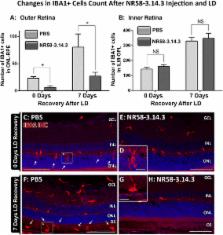
          We report that intravitreal administration of NR58-3.14.3 reduces the accumulation of macrophages in the outer retina following exposure to light damage, at both 0- and 7-day post-exposure time points. Injection of NR58-3.14.3 also reduced the up-regulation of inflammatory markers including of Il6, Ccl3, and Ccl4 in infiltrating macrophages, which are promoters of their pathogenic activity in the retina. Finally, NR58-3.14.3-injected retinas displayed markedly reduced photoreceptor death following light damage, at both 0 and 7 days post-exposure.

          Conclusions

          Our findings indicate that NR58-3.14.3 is effective in inhibiting subretinal macrophage accumulation in light-induced retinal degeneration and illustrate the potential of broad-spectrum chemokine inhibitors as novel therapeutic agents in thwarting retinal inflammation. Although broad-spectrum chemokine inhibitors may not be appropriate for all retinal inflammatory conditions, our results suggest that they may be beneficial for retinal dystrophies in which chemokine expression and subretinal macrophage accumulation are implicated, such as advanced AMD.

          Related collections

          Most cited references57

          • Record: found
          • Abstract: found
          • Article: not found

          The pivotal role of the complement system in aging and age-related macular degeneration: hypothesis re-visited.

          During the past ten years, dramatic advances have been made in unraveling the biological bases of age-related macular degeneration (AMD), the most common cause of irreversible blindness in western populations. In that timeframe, two distinct lines of evidence emerged which implicated chronic local inflammation and activation of the complement cascade in AMD pathogenesis. First, a number of complement system proteins, complement activators, and complement regulatory proteins were identified as molecular constituents of drusen, the hallmark extracellular deposits associated with early AMD. Subsequently, genetic studies revealed highly significant statistical associations between AMD and variants of several complement pathway-associated genes including: Complement factor H (CFH), complement factor H-related 1 and 3 (CFHR1 and CFHR3), complement factor B (CFB), complement component 2 (C2), and complement component 3 (C3). In this article, we revisit our original hypothesis that chronic local inflammatory and immune-mediated events at the level of Bruch's membrane play critical roles in drusen biogenesis and, by extension, in the pathobiology of AMD. Secondly, we report the results of a new screening for additional AMD-associated polymorphisms in a battery of 63 complement-related genes. Third, we identify and characterize the local complement system in the RPE-choroid complex - thus adding a new dimension of biological complexity to the role of the complement system in ocular aging and AMD. Finally, we evaluate the most salient, recent evidence that bears directly on the role of complement in AMD pathogenesis and progression. Collectively, these recent findings strongly re-affirm the importance of the complement system in AMD. They lay the groundwork for further studies that may lead to the identification of a transcriptional disease signature of AMD, and hasten the development of new therapeutic approaches that will restore the complement-modulating activity that appears to be compromised in genetically susceptible individuals. Copyright 2009 Elsevier Ltd. All rights reserved.
            Bookmark
            • Record: found
            • Abstract: found
            • Article: not found

            Retinal microglia: just bystander or target for therapy?

            Resident microglial cells can be regarded as the immunological watchdogs of the brain and the retina. They are active sensors of their neuronal microenvironment and rapidly respond to various insults with a morphological and functional transformation into reactive phagocytes. There is strong evidence from animal models and in situ analyses of human tissue that microglial reactivity is a common hallmark of various retinal degenerative and inflammatory diseases. These include rare hereditary retinopathies such as retinitis pigmentosa and X-linked juvenile retinoschisis but also comprise more common multifactorial retinal diseases such as age-related macular degeneration, diabetic retinopathy, glaucoma, and uveitis as well as neurological disorders with ocular manifestation. In this review, we describe how microglial function is kept in balance under normal conditions by cross-talk with other retinal cells and summarize how microglia respond to different forms of retinal injury. In addition, we present the concept that microglia play a key role in local regulation of complement in the retina and specify aspects of microglial aging relevant for chronic inflammatory processes in the retina. We conclude that this resident immune cell of the retina cannot be simply regarded as bystander of disease but may instead be a potential therapeutic target to be modulated in the treatment of degenerative and inflammatory diseases of the retina.
              Bookmark
              • Record: found
              • Abstract: found
              • Article: not found

              Minocycline reduces proinflammatory cytokine expression, microglial activation, and caspase-3 activation in a rodent model of diabetic retinopathy.

              Diabetes leads to vascular leakage, glial dysfunction, and neuronal apoptosis within the retina. The goal of the studies reported here was to determine the role that retinal microglial cells play in diabetic retinopathy and assess whether minocycline can decrease microglial activation and alleviate retinal complications. Immunohistochemical analyses showed that retinal microglia are activated early in diabetes. Furthermore, mRNAs for interleukin-1beta and tumor necrosis factor-alpha, proinflammatory mediators known to be released from microglia, are also increased in the retina early in the course of diabetes. Using an in vitro bioassay, we demonstrated that cytokine-activated microglia release cytotoxins that kill retinal neurons. Furthermore, we showed that neuronal apoptosis is increased in the diabetic retina, as measured by caspase-3 activity. Minocycline represses diabetes-induced inflammatory cytokine production, reduces the release of cytotoxins from activated microglia, and significantly reduces measurable caspase-3 activity within the retina. These results indicate that inhibiting microglial activity may be an important strategy in the treatment of diabetic retinopathy and that drugs such as minocycline hold promise in delaying or preventing the loss of vision associated with this disease.
                Bookmark

                Author and article information

                Contributors
                matt.rutar@anu.edu.au
                Journal
                J Neuroinflammation
                J Neuroinflammation
                Journal of Neuroinflammation
                BioMed Central (London )
                1742-2094
                24 February 2016
                24 February 2016
                2016
                : 13
                : 47
                Affiliations
                [ ]The John Curtin School of Medical Research, The Australian National University, Building 131, Garran Rd, Canberra, ACT 2601 Australia
                [ ]ANU Medical School, The Australian National University, Canberra, Australia
                Article
                514
                10.1186/s12974-016-0514-x
                4765229
                26911327
                5d06cfc0-c533-4760-9504-eaba2a8a27be
                © Fernando et al. 2016

                Open AccessThis article is distributed under the terms of the Creative Commons Attribution 4.0 International License ( http://creativecommons.org/licenses/by/4.0/), which permits unrestricted use, distribution, and reproduction in any medium, provided you give appropriate credit to the original author(s) and the source, provide a link to the Creative Commons license, and indicate if changes were made. The Creative Commons Public Domain Dedication waiver ( http://creativecommons.org/publicdomain/zero/1.0/) applies to the data made available in this article, unless otherwise stated.

                History
                : 11 December 2015
                : 18 February 2016
                Funding
                Funded by: FundRef http://dx.doi.org/10.13039/501100001108, The Ophthalmic Research Institute of Australia;
                Award ID: New Investigator Grant
                Award Recipient :
                Funded by: FundRef http://dx.doi.org/10.13039/501100000925, National Health and Medical Research Council (AU);
                Award ID: Project Grant
                Award Recipient :
                Categories
                Research
                Custom metadata
                © The Author(s) 2016

                Neurosciences
                Neurosciences

                Comments

                Comment on this article