2
views
0
recommends
+1 Recommend
0 collections
    0
    shares
      • Record: found
      • Abstract: found
      • Article: found
      Is Open Access

      STING-dependent paracriny shapes apoptotic priming of breast tumors in response to anti-mitotic treatment

      research-article

      Read this article at

      Bookmark
          There is no author summary for this article yet. Authors can add summaries to their articles on ScienceOpen to make them more accessible to a non-specialist audience.

          Abstract

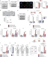
          A fascinating but uncharacterized action of antimitotic chemotherapy is to collectively prime cancer cells to apoptotic mitochondrial outer membrane permeabilization (MOMP), while impacting only on cycling cell subsets. Here, we show that a proapoptotic secretory phenotype is induced by activation of cGAS/STING in cancer cells that are hit by antimitotic treatment, accumulate micronuclei and maintain mitochondrial integrity despite intrinsic apoptotic pressure. Organotypic cultures of primary human breast tumors and patient-derived xenografts sensitive to paclitaxel exhibit gene expression signatures typical of type I IFN and TNFα exposure. These cytokines induced by cGAS/STING activation trigger NOXA expression in neighboring cells and render them acutely sensitive to BCL-xL inhibition. cGAS/STING-dependent apoptotic effects are required for paclitaxel response in vivo, and they are amplified by sequential, but not synchronous, administration of BH3 mimetics. Thus anti-mitotic agents propagate apoptotic priming across heterogeneously sensitive cancer cells through cytosolic DNA sensing pathway-dependent extracellular signals, exploitable by delayed MOMP targeting.

          Abstract

          Antimitotic compounds, such as paclitaxel, induce cell death in cycling cancer cells only. Here, the authors show that paclitaxel-targeted breast cancer cells prime neighboring cells to apoptosis through a STING-mediated paracrine signaling pathway.

          Related collections

          Most cited references37

          • Record: found
          • Abstract: found
          • Article: not found

          PARP Inhibitor Efficacy Depends on CD8+ T-cell Recruitment via Intratumoral STING Pathway Activation in BRCA-Deficient Models of Triple-Negative Breast Cancer

          Combinatorial clinical trials of PARP inhibitors with immunotherapies are ongoing, yet the immunomodulatory effects of PARP inhibition have been incompletely studied. Here, we sought to dissect the mechanisms underlying PARP inhibitor-induced changes in the tumor microenvironment of BRCA1-deficient triple-negative breast cancer (TNBC). We demonstrate that the PARP inhibitor olaparib induces CD8 + T cell infiltration and activation in vivo , and that CD8 + T cell depletion severely compromises anti-tumor efficacy. Olaparib-induced T cell recruitment is mediated through activation of the cGAS/STING pathway in tumor cells with paracrine activation of dendritic cells and is more pronounced in HR-deficient compared to HR-proficient TNBC cells and in vivo models. CRISPR-knockout of STING in cancer cells prevents proinflammatory signaling and is sufficient to abolish olaparib-induced T cell infiltration in vivo . These findings elucidate an additional mechanism of action of PARP inhibitors and provide rationale for combining PARP inhibition with immunotherapies for the treatment of TNBC.
            Bookmark
            • Record: found
            • Abstract: found
            • Article: not found

            Phase I study of Navitoclax (ABT-263), a novel Bcl-2 family inhibitor, in patients with small-cell lung cancer and other solid tumors.

            Resistance to chemotherapy-induced apoptosis represents a major obstacle to cancer control. Overexpression of Bcl-2 is seen in multiple tumor types and targeting Bcl-2 may provide therapeutic benefit. A phase I study of navitoclax, a novel inhibitor of Bcl-2 family proteins, was conducted to evaluate safety, pharmacokinetics, and preliminary efficacy in patients with solid tumors. Patients enrolled to intermittent dosing cohorts received navitoclax on day -3, followed by dosing on days 1 to 14 of a 21-day cycle. Patients on continuous dosing received a 1-week lead-in dose of 150 mg followed by continuous daily administration. Blood samples were collected for pharmacokinetic analyses, biomarker analyses, and platelet monitoring. Forty-seven patients, including 29 with small-cell lung cancer (SCLC) or pulmonary carcinoid, were enrolled between 2007 and 2008, 35 on intermittent and 12 on continuous dosing cohorts. Primary toxicities included diarrhea (40%), nausea (34%), vomiting (36%), and fatigue (34%); most were grade 1 or 2. Dose- and schedule-dependent thrombocytopenia was seen in all patients. One patient with SCLC had a confirmed partial response lasting longer than 2 years, and eight patients with SCLC or carcinoid had stable disease (one remained on study for 13 months). Pro-gastrin releasing peptide (pro-GRP) was identified as a surrogate marker of Bcl-2 amplification and changes correlated with changes in tumor volume. Navitoclax is safe and well tolerated, with dose-dependent thrombocytopenia as the major adverse effect. Preliminary efficacy data are encouraging in SCLC. Efficacy in SCLC and the utility of pro-GRP as a marker of treatment response will be further evaluated in phase II studies.
              Bookmark
              • Record: found
              • Abstract: found
              • Article: not found

              Drug-induced death signaling strategy rapidly predicts cancer response to chemotherapy.

              There is a lack of effective predictive biomarkers to precisely assign optimal therapy to cancer patients. While most efforts are directed at inferring drug response phenotype based on genotype, there is very focused and useful phenotypic information to be gained from directly perturbing the patient's living cancer cell with the drug(s) in question. To satisfy this unmet need, we developed the Dynamic BH3 Profiling technique to measure early changes in net pro-apoptotic signaling at the mitochondrion ("priming") induced by chemotherapeutic agents in cancer cells, not requiring prolonged ex vivo culture. We find in cell line and clinical experiments that early drug-induced death signaling measured by Dynamic BH3 Profiling predicts chemotherapy response across many cancer types and many agents, including combinations of chemotherapies. We propose that Dynamic BH3 Profiling can be used as a broadly applicable predictive biomarker to predict cytotoxic response of cancers to chemotherapeutics in vivo.
                Bookmark

                Author and article information

                Contributors
                philippe.juin@univ-nantes.fr
                sophie.barille@inserm.fr
                Journal
                Nat Commun
                Nat Commun
                Nature Communications
                Nature Publishing Group UK (London )
                2041-1723
                14 January 2020
                14 January 2020
                2020
                : 11
                : 259
                Affiliations
                [1 ]GRID grid.4817.a, CRCINA, INSERM, Université d’Angers, Université de Nantes, ; Nantes, France
                [2 ]SIRIC ILIAD, Nantes, Angers, France
                [3 ]ISNI 0000 0000 9437 3027, GRID grid.418191.4, Institut de Cancérologie de l’Ouest, ; 15 Rue André Boquel, 49055 Angers, Pays de la Loire France
                [4 ]ISNI 0000 0001 2175 3974, GRID grid.418682.1, Oniris, site Chantrerie, CS40706, ; 44307 Cedex 3 Nantes, France
                [5 ]UMR 1280 PhAN, Université de Nantes, INRA, Nantes, France
                [6 ]Nantes Université, CHU Nantes, Inserm, CRTI, UMR 1064, ITUN, Nantes, France
                [7 ]Nantes Université, CHU Nantes, Inserm, CNRS, SFR Santé, FED 4203, Inserm UMS 016, CNRS UMS 3556 Nantes, France
                [8 ]ISNI 0000 0004 0472 0371, GRID grid.277151.7, Service d’Anatomie Pathologique, CHU Nantes, ; Nantes, France
                Author information
                http://orcid.org/0000-0001-7203-2982
                http://orcid.org/0000-0003-3594-0353
                http://orcid.org/0000-0002-4997-3888
                http://orcid.org/0000-0001-5171-9937
                Article
                13689
                10.1038/s41467-019-13689-y
                6959316
                31937780
                5d11a94b-aeac-4d89-bd84-99fc1b73f976
                © The Author(s) 2020

                Open Access This article is licensed under a Creative Commons Attribution 4.0 International License, which permits use, sharing, adaptation, distribution and reproduction in any medium or format, as long as you give appropriate credit to the original author(s) and the source, provide a link to the Creative Commons license, and indicate if changes were made. The images or other third party material in this article are included in the article’s Creative Commons license, unless indicated otherwise in a credit line to the material. If material is not included in the article’s Creative Commons license and your intended use is not permitted by statutory regulation or exceeds the permitted use, you will need to obtain permission directly from the copyright holder. To view a copy of this license, visit http://creativecommons.org/licenses/by/4.0/.

                History
                : 25 February 2019
                : 21 November 2019
                Funding
                Funded by: Ligue contre le Cancer Grand-Ouest (44, 85, 53, 56, 22)
                Categories
                Article
                Custom metadata
                © The Author(s) 2020

                Uncategorized
                cancer,breast cancer,cell biology,cell death,apoptosis
                Uncategorized
                cancer, breast cancer, cell biology, cell death, apoptosis

                Comments

                Comment on this article