16
views
0
recommends
+1 Recommend
0 collections
    0
    shares
      • Record: found
      • Abstract: found
      • Article: found
      Is Open Access

      Pulmonary embolism presenting with itinerant chest pain and migratory pleural effusion : A case report

      case-report

      Read this article at

      Bookmark
          There is no author summary for this article yet. Authors can add summaries to their articles on ScienceOpen to make them more accessible to a non-specialist audience.

          Abstract

          Introduction:

          Pulmonary embolism (PE) presents with complex clinical manifestations ranging from asymptomatic to chest pain, hemoptysis, syncope, shock, or sudden death. To the authors’ knowledge, itinerant chest pain has not been reported as sign or symptom of PE.

          Case presentation:

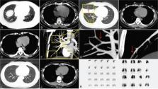
          A 41-year-old woman presenting with left chest pain, no hemoptysis, or breathing difficulties. The chest pain was more severe on deep inspiration. Chest computed tomography (CT) and ultrasound imaging showed left pleural effusion. After antibiotic treatment, the left chest pain was alleviated, but a similar pain appeared in the right chest. Electrocardiogram, blood gas analysis, echocardiography, and D-dimer levels were unremarkable. Chest CT showed right pleural effusion. A CT pulmonary angiography (CTPA) unexpectedly revealed a PE in the right pulmonary artery. The patient was administered anticoagulant therapy and made a complete recovery.

          Conclusions:

          The use of CTPA to investigate the possible presence of PE in patients with unexplained migratory pleural effusion complaining of itinerant chest pain is important. Lessons should be learned from the early use of CTPA to investigate the possible presence of PE in patients.

          Related collections

          Most cited references19

          • Record: found
          • Abstract: found
          • Article: not found

          Derivation of a simple clinical model to categorize patients probability of pulmonary embolism: increasing the models utility with the SimpliRED D-dimer.

          We have previously demonstrated that a clinical model can be safely used in a management strategy in patients with suspected pulmonary embolism (PE). We sought to simplify the clinical model and determine a scoring system, that when combined with D-dimer results, would safely exclude PE without the need for other tests, in a large proportion of patients. We used a randomly selected sample of 80% of the patients that participated in a prospective cohort study of patients with suspected PE to perform a logistic regression analysis on 40 clinical variables to create a simple clinical prediction rule. Cut points on the new rule were determined to create two scoring systems. In the first scoring system patients were classified as having low, moderate and high probability of PE with the proportions being similar to those determined in our original study. The second system was designed to create two categories, PE likely and unlikely. The goal in the latter was that PE unlikely patients with a negative D-dimer result would have PE in less than 2% of cases. The proportion of patients with PE in each category was determined overall and according to a positive or negative SimpliRED D-dimer result. After these determinations we applied the models to the remaining 20% of patients as a validation of the results. The following seven variables and assigned scores (in brackets) were included in the clinical prediction rule: Clinical symptoms of DVT (3.0), no alternative diagnosis (3.0), heart rate >100 (1.5), immobilization or surgery in the previous four weeks (1.5), previous DVT/PE (1.5), hemoptysis (1.0) and malignancy (1.0). Patients were considered low probability if the score was 4.0. 7.8% of patients with scores of less than or equal to 4 had PE but if the D-dimer was negative in these patients the rate of PE was only 2.2% (95% CI = 1.0% to 4.0%) in the derivation set and 1.7% in the validation set. Importantly this combination occurred in 46% of our study patients. A score of <2.0 and a negative D-dimer results in a PE rate of 1.5% (95% CI = 0.4% to 3.7%) in the derivation set and 2.7% (95% CI = 0.3% to 9.0%) in the validation set and only occurred in 29% of patients. The combination of a score < or =4.0 by our simple clinical prediction rule and a negative SimpliRED D-Dimer result may safely exclude PE in a large proportion of patients with suspected PE.
            Bookmark
            • Record: found
            • Abstract: found
            • Article: not found

            The diagnosis of pleural effusions.

            Pleural effusions arise from a variety of systemic, inflammatory, infectious and malignant conditions. Their precise etiological diagnosis depends on a combination of medical history, physical examination, imaging tests and pertinent pleural fluid analyses; including specific biomarkers (e.g., natriuretic peptides for heart failure, adenosine deaminase for tuberculosis, or mesothelin for mesothelioma). Invasive procedures, such as pleuroscopic biopsies, may be required for persistently symptomatic effusions which remain undiagnosed after the analysis of one or more pleural fluid samples. However, whenever parietal pleural nodularity or thickening exist, image-guided biopsies should first be attempted. This review addresses the current diagnostic approach to pleural effusions secondary to heart failure, pneumonia, cancer, tuberculosis and other less frequent conditions.
              Bookmark
              • Record: found
              • Abstract: found
              • Article: not found

              Parenchymal and pleural findings in patients with and patients without acute pulmonary embolism detected at spiral CT.

              To compare the frequencies of parenchymal abnormalities and pleural effusions in patients with and patients without acute pulmonary embolism (PE) detected at spiral computed tomography (CT). Contrast material-enhanced spiral CT scans obtained in 92 patients clinically suspected of having acute PE were retrospectively reviewed. The presence or absence of parenchymal abnormalities and pleural effusions was noted. The presence of filling defects consistent with central or peripheral PE was recorded. Twenty-eight patients had CT evidence of PE. Central emboli were evident in 27 (96%) of these patients; 23 (82%) had concomitant central and peripheral emboli, and four (14%) had only central emboli. One patient had an isolated subsegmental clot. Parenchymal abnormalities were seen in 24 (86%) patients with PE and 56 (88%) patients without PE. Atelectasis, the most common finding, was present in 20 (71%) patients with PE and 41 (64%) patients without PE. The only parenchymal abnormality significantly associated with PE was peripheral wedge-shaped opacity, which was seen in seven (25%) patients with PE and three (5%) patients without PE (odds ratio, 6.78; 95% CI = 1.60, 28.62). Pleural effusions were seen in 16 (57%) patients with PE and 36 (56%) patients without PE. In 25 (39%) patients without PE, there were additional CT findings that might suggest an alternative explanation for the acute clinical presentation. Parenchymal and pleural findings at CT are of limited value for differentiating patients with PE from those without PE.
                Bookmark

                Author and article information

                Journal
                Medicine (Baltimore)
                Medicine (Baltimore)
                MEDI
                Medicine
                Wolters Kluwer Health
                0025-7974
                1536-5964
                June 2018
                01 June 2018
                : 97
                : 22
                : e10944
                Affiliations
                [a ]Department of Respiratory and Critical Care Medicine, The Second Hospital of Jilin University, Changchun, Jilin
                [b ]Department of Respiratory, The Fourth Hospital of Daqing City, DaQing, Helongjiang, China.
                Author notes
                []Correspondence: Peng Gao, Department of Respiratory and Critical Care Medicine, The Second Hospital of Jilin University, Changchun, Jilin 130041, China (e-mail: gaopeng1234@ 123456sina.com )
                Article
                MD-D-18-01404 10944
                10.1097/MD.0000000000010944
                6392842
                29851836
                5f671a0a-085e-4199-a80b-2ba2970f5fae
                Copyright © 2018 the Author(s). Published by Wolters Kluwer Health, Inc.

                This is an open access article distributed under the Creative Commons Attribution License 4.0 (CCBY), which permits unrestricted use, distribution, and reproduction in any medium, provided the original work is properly cited. http://creativecommons.org/licenses/by/4.0

                History
                : 28 February 2018
                : 8 May 2018
                Categories
                6700
                Research Article
                Clinical Case Report
                Custom metadata
                TRUE

                chest pain,computed tomographic pulmonary angiography,d-dimer,pulmonary embolism,pleural effusion

                Comments

                Comment on this article