11
views
0
recommends
+1 Recommend
0 collections
    0
    shares
      • Record: found
      • Abstract: found
      • Article: found
      Is Open Access

      Echocardiographic characteristics of primary malignant pericardial mesothelioma and outcomes analysis: a retrospective study

      research-article

      Read this article at

      Bookmark
          There is no author summary for this article yet. Authors can add summaries to their articles on ScienceOpen to make them more accessible to a non-specialist audience.

          Abstract

          Background

          Little is known about the echocardiographic characteristics of primary malignant pericardial mesothelioma (PPM) due to its rarity. The aim of this study was to explore the sex-specific echocardiographic patterns of PPM and risk factors for in-hospital mortality.

          Methods

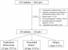
          A retrospective information retrieval was conducted for cases of PPM reported from China during 1981 and 2015. The diagnosis was made by histopathological examinations and only cases with echocardiographic descriptions were included. Data on the clinical and echocardiographic findings were collected. Difference in clinical, sex-specific echocardiographic characteristics and findings across different time periods were assessed. Logistic regression analysis was performed to explore echocardiographic risk factors for in-hospital mortality.

          Results

          A total of 64 patients with PPM were included, with a mean age of 39.2 ± 15.6 years and minor male dominance (40, 62.5%). The most common echocardiographic presentations were pericardial effusion (55, 85.9%), pericardial masses (36.4%) and thickening (17.3%), respectively. The positive rate of pericardiocentesis was only 20.9%. Six patients (15.4%) died among 39 cases reporting in-hospital outcome. Logistics analysis identified no clinical or echocardiographic parameters associated with in-hospital mortality (all P > 0.05).

          Conclusions

          The echocardiographic signs of PPM are basically nonspecific with massive pericardial effusion as the most common sign, although no echocardiographic gender differences or association with in-hospital mortality could be identified.

          Related collections

          Most cited references19

          • Record: found
          • Abstract: found
          • Article: not found

          Tumors of the heart. A 20-year experience with a review of 12,485 consecutive autopsies.

          Cardiac involvement by primary and secondary tumors is one of the least investigated subjects in oncology. Seven cases of primary and 154 cases of secondary cardiac tumors from autopsies performed over a 20-year period (1972 through 1991) at Queen Mary Hospital, Hong Kong, were reviewed. During this period, 12,485 autopsies were performed, and the autopsy incidence for primary and secondary heart tumors is thus 0.056% and 1.23%, respectively. Only seven primary cardiac tumors were found, including two myxomas, two rhabdomyomas, two hemangiomas, and one lipoma. For secondary tumors involving the heart (including both metastasis and local extension), important primary tumors in male subjects were carcinoma of the lung (31.7%), esophageal carcinoma (28.7%), lymphoma (11.9%), carcinoma of the liver (6.9%), leukemia (4.0%), and gastric carcinoma (4.0%), while in female subjects, carcinoma of the lung (35.9%), lymphoma (17.0%), carcinoma of the breast (7.5%), and pancreatic carcinoma (7.5%) predominated. Overall, the three most common malignant neoplasms encountered were carcinoma of the lung, esophageal carcinoma, and lymphoma. Pericardium, including epicardium, was the most common location of cardiac involvement by secondary tumors, followed by myocardium and endocardium. The present study showed a higher percentage of esophageal carcinoma and carcinoma of the liver (reflecting the higher incidence of these tumors in Hong Kong Chinese), but a lower incidence of carcinoma of the breast when compared with other series. The metastatic lung tumors showed an unusual predominance of adenocarcinoma.
            Bookmark
            • Record: found
            • Abstract: found
            • Article: not found

            Left ventricular pseudoaneurysm.

            Left ventricular (LV) pseudoaneurysms form when cardiac rupture is contained by adherent pericardium or scar tissue. Although LV pseudoaneurysms are not common, the diagnosis is difficult and they are prone to rupture. We evaluated the clinical presentation, diagnostic accuracy of imaging modalities, results of therapy and prognosis of 290 patients with LV pseudoaneurysms. Most cases of LV pseudoaneurysm were related to myocardial infarction (particularly inferior wall myocardial infarction) and cardiac surgery. Congestive heart failure, chest pain and dyspnea were the most frequently reported symptoms, but >10% of patients were asymptomatic. Physical examination revealed a murmur in 70% of patients. Almost all patients had electrocardiographic abnormalities, but these were usually nonspecific ST segment changes; only 20% of patients had ST segment elevation. Although radiographic findings were also usually nonspecific, the appearance of a mass was present in more than one half of patients and may be an important clue to the correct diagnosis. Left ventricular angiography was the most definitive test and can be useful in planning surgery since concomitant coronary angiography can be performed. Regardless of treatment, patients with LV pseudoaneurysms had a high mortality rate, especially those who did not undergo surgery. Because the symptoms, signs, electrocardiographic abnormalities and radiographic findings seen in patients with LV pseudoaneurysms can be indistinguishable from those in patients with coronary disease alone, a high clinical index of suspicion is needed to avoid missing the diagnosis.
              Bookmark
              • Record: found
              • Abstract: found
              • Article: not found

              Cardiac tumors in a tertiary care cancer hospital: clinical features, echocardiographic findings, treatment and outcomes

              Cardiac tumors are a rare entity, comprised of tumors with diverse histology and natural history. We report the clinical characteristics, echocardiograhic findings, therapy and outcome of 59 patients with primary and metastatic cardiac tumors. Our institutional echocardiogram data base from 1993 through 2005 was reviewed to identify patients diagnosed with intra-cardiac tumor. A total of 59 patients with cardiac tumors were identified and included in the study. The patients' characteristics, presenting symptoms, diagnostic tests, location, histology of the tumor, treatment and the one year survival rate of this population was collected from the medical records. Of the 59 cardiac tumor cases, 16 (27%) were primary cardiac tumors and 43 (73%) were secondary cardiac tumors. The most common primary tumor was sarcoma affecting 13 (81%) of the 16 cases. Of these, 5 patients were angiosarcoma, 5 unclassified sarcoma, one myxoid sarcoma and 2 malignant fibrous histiocytoma. The mean age at presentation was 41.1 years, and the most common location was right atrium affecting 6 cases (37.5%). The most common symptom of dyspnea was present in 10 (62.5%) cases. Eleven (25.6%) of the 43 secondary cardiac tumors were metastasis from renal cell carcinoma. The mean age at presentation was 55.4 years. Right atrium was the most frequent location affecting 18 (42%) of the 43 patients. The most common presenting symptom was dyspnea in 15 (35%) cases. For both primary and secondary tumors, dyspnea was the most common symptom and right atrium was most frequently involved. Sarcoma was the most common primary cardiac tumor while metastasis from renal cell carcinoma was the most common secondary tumor.
                Bookmark

                Author and article information

                Contributors
                +86 010 85231403 , lvxzcyh@163.com
                Journal
                Cardiovasc Ultrasound
                Cardiovasc Ultrasound
                Cardiovascular Ultrasound
                BioMed Central (London )
                1476-7120
                26 April 2018
                26 April 2018
                2018
                : 16
                : 7
                Affiliations
                [1 ]ISNI 0000 0004 0369 153X, GRID grid.24696.3f, Echocardiography Department of Heart Center, Beijing Chao-Yang Hospital, , Capital Medical University, ; 8 Gongren Tiyuchang Nanlu, Chaoyang District, Beijing, 100020 China
                [2 ]Department of Cardiology, Jiang Xi Yichun Hospital of Traditional Chinese Medicine, Jiang Xi, China
                [3 ]ISNI 0000 0004 0632 4559, GRID grid.411634.5, Department of Cardiology, , Beijing Daxing District people’s Hospital, ; Beijing, China
                Article
                125
                10.1186/s12947-018-0125-z
                5922299
                29695235
                60314ca0-9220-4fa5-a6cf-b463830f0f9d
                © The Author(s). 2018

                Open Access This article is distributed under the terms of the Creative Commons Attribution 4.0 International License ( http://creativecommons.org/licenses/by/4.0/), which permits unrestricted use, distribution, and reproduction in any medium, provided you give appropriate credit to the original author(s) and the source, provide a link to the Creative Commons license, and indicate if changes were made. The Creative Commons Public Domain Dedication waiver ( http://creativecommons.org/publicdomain/zero/1.0/) applies to the data made available in this article, unless otherwise stated.

                History
                : 11 December 2017
                : 4 April 2018
                Categories
                Research
                Custom metadata
                © The Author(s) 2018

                Cardiovascular Medicine
                pericardial mesothelioma,echocardiography,diagnosis,prognosis
                Cardiovascular Medicine
                pericardial mesothelioma, echocardiography, diagnosis, prognosis

                Comments

                Comment on this article