635
views
0
recommends
+1 Recommend
0 collections
    20
    shares
      • Record: found
      • Abstract: found
      • Article: found
      Is Open Access

      Development of SNAP-Tag Fluorogenic Probes for Wash-Free Fluorescence Imaging

      research-article

      Read this article at
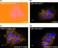
      Bookmark
          There is no author summary for this article yet. Authors can add summaries to their articles on ScienceOpen to make them more accessible to a non-specialist audience.

          Abstract

          The ability to specifically attach chemical probes to individual proteins represents a powerful approach to the study and manipulation of protein function in living cells. It provides a simple, robust and versatile approach to the imaging of fusion proteins in a wide range of experimental settings. However, a potential drawback of detection using chemical probes is the fluorescence background from unreacted or nonspecifically bound probes. In this report we present the design and application of novel fluorogenic probes for labeling SNAP-tag fusion proteins in living cells. SNAP-tag is an engineered variant of the human repair protein O 6-alkylguanine-DNA alkyltransferase (hAGT) that covalently reacts with benzylguanine derivatives. Reporter groups attached to the benzyl moiety become covalently attached to the SNAP tag while the guanine acts as a leaving group. Incorporation of a quencher on the guanine group ensures that the benzylguanine probe becomes highly fluorescent only upon labeling of the SNAP-tag protein. We describe the use of intramolecularly quenched probes for wash-free labeling of cell surface-localized epidermal growth factor receptor (EGFR) fused to SNAP-tag and for direct quantification of SNAP-tagged β-tubulin in cell lysates. In addition, we have characterized a fast-labeling variant of SNAP-tag, termed SNAP f, which displays up to a tenfold increase in its reactivity towards benzylguanine substrates. The presented data demonstrate that the combination of SNAP f and the fluorogenic substrates greatly reduces the background fluorescence for labeling and imaging applications. This approach enables highly sensitive spatiotemporal investigation of protein dynamics in living cells.

          Related collections

          Most cited references42

          • Record: found
          • Abstract: found
          • Article: not found

          The fluorescent toolbox for assessing protein location and function.

          Advances in molecular biology, organic chemistry, and materials science have recently created several new classes of fluorescent probes for imaging in cell biology. Here we review the characteristic benefits and limitations of fluorescent probes to study proteins. The focus is on protein detection in live versus fixed cells: determination of protein expression, localization, activity state, and the possibility for combination of fluorescent light microscopy with electron microscopy. Small organic fluorescent dyes, nanocrystals ("quantum dots"), autofluorescent proteins, small genetic encoded tags that can be complexed with fluorochromes, and combinations of these probes are highlighted.
            Bookmark
            • Record: found
            • Abstract: found
            • Article: not found

            A general method for the covalent labeling of fusion proteins with small molecules in vivo.

            Characterizing the movement, interactions, and chemical microenvironment of a protein inside the living cell is crucial to a detailed understanding of its function. Most strategies aimed at realizing this objective are based on genetically fusing the protein of interest to a reporter protein that monitors changes in the environment of the coupled protein. Examples include fusions with fluorescent proteins, the yeast two-hybrid system, and split ubiquitin. However, these techniques have various limitations, and considerable effort is being devoted to specific labeling of proteins in vivo with small synthetic molecules capable of probing and modulating their function. These approaches are currently based on the noncovalent binding of a small molecule to a protein, the formation of stable complexes between biarsenical compounds and peptides containing cysteines, or the use of biotin acceptor domains. Here we describe a general method for the covalent labeling of fusion proteins in vivo that complements existing methods for noncovalent labeling of proteins and that may open up new ways of studying proteins in living cells.
              Bookmark
              • Record: found
              • Abstract: found
              • Article: not found

              An engineered protein tag for multiprotein labeling in living cells.

              The visualization of complex cellular processes involving multiple proteins requires the use of spectroscopically distinguishable fluorescent reporters. We have previously introduced the SNAP-tag as a general tool for the specific labeling of SNAP-tag fusion proteins in living cells. The SNAP-tag is derived from the human DNA repair protein O6-alkylguanine-DNA alkyltransferase (AGT) and can be covalently labeled in living cells using O6-benzylguanine derivatives bearing a chemical probe. Here we report the generation of an AGT-based tag, named CLIP-tag, which reacts specifically with O2-benzylcytosine derivatives. Because SNAP-tag and CLIP-tag possess orthogonal substrate specificities, SNAP and CLIP fusion proteins can be labeled simultaneously and specifically with different molecular probes in living cells. We furthermore show simultaneous pulse-chase experiments to visualize different generations of two different proteins in one sample.
                Bookmark

                Author and article information

                Journal
                Chembiochem
                cbic
                Chembiochem
                WILEY-VCH Verlag
                1439-4227
                1439-7633
                19 September 2011
                26 July 2011
                : 12
                : 14
                : 2217-2226
                Affiliations
                [[a] ]simpleNew England Biolabs, Inc 240 County Road, Ipswich, MA 01938 (USA)
                [[b] ]simpleInstitute of Chemical Sciences and Engineering, Ecole Polytechnique Federal de Lausanne (EPFL) 1015 Lausanne (Switzerland)
                Author notes
                Dr. X. Sun, Dr. A. Zhang, B. Baker, Dr. L. Sun, A. Howard, J. Buswell, Dr. C. J. Noren, Dr. M.-Q. Xu, Dr. I. R. Corrêa, Jr. New England Biolabs, Inc. 240 County Road, Ipswich, MA 01938 (USA) E-mail: correa@ 123456neb.com xum@ 123456neb.com

                Supporting information for this article is available on the WWW under http://dx.doi.org/10.1002/cbic.201100173.

                Article
                10.1002/cbic.201100173
                3213346
                21793150
                64437626-3b96-4b07-93a5-6b3d7439c06c
                Copyright © 2011 WILEY-VCH Verlag GmbH & Co. KGaA, Weinheim

                Re-use of this article is permitted in accordance with the Creative Commons Deed, Attribution 2.5, which does not permit commercial exploitation.

                History
                : 15 March 2011
                Categories
                Full Paper

                Biochemistry
                fluorogenic substrates,covalent labeling,protein modifications,cell imaging,fluorescent probes

                Comments

                Comment on this article