13
views
0
recommends
+1 Recommend
0 collections
    0
    shares
      • Record: found
      • Abstract: found
      • Article: found
      Is Open Access

      Endometrium and endometriosis tissue mitochondrial energy metabolism in a nonhuman primate model

      research-article

      Read this article at

      Bookmark
          There is no author summary for this article yet. Authors can add summaries to their articles on ScienceOpen to make them more accessible to a non-specialist audience.

          Abstract

          Background

          Endometriosis is the growth of uterine lining (endometrium) outside of the uterus. In other chronic inflammatory diseases, mitochondrial dysfunction is suspected of playing a role in disease pathogenesis. However, little is known about endometriosis mitochondrial function or its effects on tissue metabolism. The objectives of this study were to analyze mitochondrial function in nonhuman primate (NHP) endometrium and endometriosis tissue and to identify the metabolic features of these tissues that may contribute to disease.

          Methods

          Mitochondrial function in endometriosis tissue and endometrium was measured using mitochondrial respirometry analysis to determine if changes in oxidative phosphorylation exist in endometrium and endometriosis tissue compared to control endometrium from clinically healthy NHPs. Targeted metabolomics and multidimensional statistical analysis were applied to quantify key metabolites in energy and amino acid biosynthesis pathways.

          Results

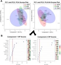
          Mitochondrial respirometry assays showed endometrium from NHPs with endometriosis had reduced complex II-mediated oxygen consumption rates (OCR) across all energy states (basal, p = 0.01; state 3, p = 0.02; state 3u, p = 0.04; state 4o, p = 0.008) and endometriosis tissue had reduced state 3, complex I-mediated OCR ( p = 0.02) and respiratory control rates ( p = 0.01) compared to normal endometrium. Targeted metabolomics performed on tissue revealed carnitine ( p = 0.001), creatine phosphate ( p = 0.01), NADH ( p = 0.0001), FAD ( p = 0.001), tryptophan ( p = 0.0009), and malic acid ( p = 0.005) were decreased in endometriosis tissue compared to normal endometrium samples. FAD ( p = 0.004), tryptophan ( p = 0.0004) and malic acid ( p = 0.03) were significantly decreased in endometrium from NHPs with endometriosis compared to normal endometrium. Significant metabolites identified in endometriosis and endometrium samples from animals with endometriosis were part of amino acid biosynthesis or energy metabolism pathways.

          Conclusions

          Here, endometrial mitochondrial energy production and metabolism were decreased in endometrium and endometriosis tissue. Decreased mitochondrial energy production may be due to oxidative stress-induced damage to mitochondrial DNA or membranes, a shift in cell metabolism, or decreased energy substrate; however, the exact cause remains unknown. Additional research is needed to determine the implications of reduced mitochondrial energy production and metabolism on endometriosis and endometrium.

          Electronic supplementary material

          The online version of this article (10.1186/s12958-019-0513-8) contains supplementary material, which is available to authorized users.

          Related collections

          Most cited references28

          • Record: found
          • Abstract: found
          • Article: found
          Is Open Access

          MetaboAnalyst: a web server for metabolomic data analysis and interpretation

          Metabolomics is a newly emerging field of ‘omics’ research that is concerned with characterizing large numbers of metabolites using NMR, chromatography and mass spectrometry. It is frequently used in biomarker identification and the metabolic profiling of cells, tissues or organisms. The data processing challenges in metabolomics are quite unique and often require specialized (or expensive) data analysis software and a detailed knowledge of cheminformatics, bioinformatics and statistics. In an effort to simplify metabolomic data analysis while at the same time improving user accessibility, we have developed a freely accessible, easy-to-use web server for metabolomic data analysis called MetaboAnalyst. Fundamentally, MetaboAnalyst is a web-based metabolomic data processing tool not unlike many of today's web-based microarray analysis packages. It accepts a variety of input data (NMR peak lists, binned spectra, MS peak lists, compound/concentration data) in a wide variety of formats. It also offers a number of options for metabolomic data processing, data normalization, multivariate statistical analysis, graphing, metabolite identification and pathway mapping. In particular, MetaboAnalyst supports such techniques as: fold change analysis, t-tests, PCA, PLS-DA, hierarchical clustering and a number of more sophisticated statistical or machine learning methods. It also employs a large library of reference spectra to facilitate compound identification from most kinds of input spectra. MetaboAnalyst guides users through a step-by-step analysis pipeline using a variety of menus, information hyperlinks and check boxes. Upon completion, the server generates a detailed report describing each method used, embedded with graphical and tabular outputs. MetaboAnalyst is capable of handling most kinds of metabolomic data and was designed to perform most of the common kinds of metabolomic data analyses. MetaboAnalyst is accessible at http://www.metaboanalyst.ca
            Bookmark
            • Record: found
            • Abstract: found
            • Article: not found

            Oxidative stress response elicited by mitochondrial dysfunction: implication in the pathophysiology of aging.

            Under normal physiological conditions, reactive oxygen species (ROS) serve as 'redox messengers' in the regulation of intracellular signalling, whereas excess ROS may induce irreversible damage to cellular components and lead to cell death by promoting the intrinsic apoptotic pathway through mitochondria. In the aging process, accumulation of mitochondria DNA mutations, impairment of oxidative phosphorylation as well as an imbalance in the expression of antioxidant enzymes result in further overproduction of ROS. This mitochondrial dysfunction-elicited ROS production axis forms a vicious cycle, which is the basis of mitochondrial free radical theory of aging. In addition, several lines of evidence have emerged recently to demonstrate that ROS play crucial roles in the regulation of cellular metabolism, antioxidant defence and posttranslational modification of proteins. We first discuss the oxidative stress responses, including metabolites redistribution and alteration of the acetylation status of proteins, in human cells with mitochondrial dysfunction and in aging. On the other hand, autophagy and mitophagy eliminate defective mitochondria and serve as a scavenger and apoptosis defender of cells in response to oxidative stress during aging. These scenarios mediate the restoration or adaptation of cells to respond to aging and age-related disorders for survival. In the natural course of aging, the homeostasis in the network of oxidative stress responses is disturbed by a progressive increase in the intracellular level of the ROS generated by defective mitochondria. Caloric restriction, which is generally thought to promote longevity, has been reported to enhance the efficiency of this network and provide multiple benefits to tissue cells. In this review, we emphasize the positive and integrative roles of mild oxidative stress elicited by mitochondria in the regulation of adaptation, anti-aging and scavenging pathway beyond their roles in the vicious cycle of mitochondrial dysfunction in the aging process.
              Bookmark
              • Record: found
              • Abstract: found
              • Article: found
              Is Open Access

              Menstrual physiology: implications for endometrial pathology and beyond

              BACKGROUND Each month the endometrium becomes inflamed, and the luminal portion is shed during menstruation. The subsequent repair is remarkable, allowing implantation to occur if fertilization takes place. Aberrations in menstrual physiology can lead to common gynaecological conditions, such as heavy or prolonged bleeding. Increased knowledge of the processes involved in menstrual physiology may also have translational benefits at other tissue sites. METHODS Pubmed and Cochrane databases were searched for all original and review articles published in English until April 2015. Search terms included ‘endometrium’, ‘menstruation’, ‘endometrial repair’, ‘endometrial regeneration’ ‘angiogenesis’, ‘inflammation’ and ‘heavy menstrual bleeding’ or ‘menorrhagia’. RESULTS Menstruation occurs naturally in very few species. Human menstruation is thought to occur as a consequence of preimplantation decidualization, conferring embryo selectivity and the ability to adapt to optimize function. We highlight how current and future study of endometrial inflammation, vascular changes and repair/regeneration will allow us to identify new therapeutic targets for common gynaecological disorders. In addition, we describe how increased knowledge of this endometrial physiology will have many translational applications at other tissue sites. We highlight the clinical applications of what we know, the key questions that remain and the scientific and medical possibilities for the future. CONCLUSIONS The study of menstruation, in both normal and abnormal scenarios, is essential for the production of novel, acceptable medical treatments for common gynaecological complaints. Furthermore, collaboration and communication with specialists in other fields could significantly advance the therapeutic potential of this dynamic tissue.
                Bookmark

                Author and article information

                Contributors
                hmatkins55@gmail.com
                mbharada@gmail.com
                aocox@wakehealth.edu
                cfurdui@wakehealth.edu
                sappt@wakehealth.edu
                336.716.4763 , dcaudell@wakehealth.edu
                Journal
                Reprod Biol Endocrinol
                Reprod. Biol. Endocrinol
                Reproductive Biology and Endocrinology : RB&E
                BioMed Central (London )
                1477-7827
                24 August 2019
                24 August 2019
                2019
                : 17
                : 70
                Affiliations
                [1 ]ISNI 0000 0001 2185 3318, GRID grid.241167.7, Department of Pathology, , Section on Comparative Medicine, Wake Forest School of Medicine, ; Winston-Salem, One Medical Center Blvd, Winston-Salem, NC 27157 USA
                [2 ]ISNI 0000 0001 2185 3318, GRID grid.241167.7, Department of Internal Medicine, , Section on Molecular Medicine, Wake Forest School of Medicine, ; Winston-Salem, NC USA
                [3 ]ISNI 0000 0001 2107 5309, GRID grid.422638.9, Agilent Technologies, Cell Analysis Division, ; Lexington, MA USA
                Article
                513
                10.1186/s12958-019-0513-8
                6708555
                31445519
                64688262-cb2c-41f2-8635-5641ddbeba73
                © The Author(s). 2019

                Open AccessThis article is distributed under the terms of the Creative Commons Attribution 4.0 International License ( http://creativecommons.org/licenses/by/4.0/), which permits unrestricted use, distribution, and reproduction in any medium, provided you give appropriate credit to the original author(s) and the source, provide a link to the Creative Commons license, and indicate if changes were made. The Creative Commons Public Domain Dedication waiver ( http://creativecommons.org/publicdomain/zero/1.0/) applies to the data made available in this article, unless otherwise stated.

                History
                : 27 March 2019
                : 16 August 2019
                Funding
                Funded by: FundRef http://dx.doi.org/10.13039/100000052, NIH Office of the Director;
                Award ID: T32OD010957-37
                Funded by: FundRef http://dx.doi.org/10.13039/100000002, National Institutes of Health;
                Award ID: R01HL087103
                Award ID: UL1TR001420
                Funded by: National Institutes of Health (US)
                Award ID: P30CA012197
                Funded by: Department of Pathology-Comparative Medicine Internal Pilot Award
                Award ID: 125-120310-00000-830086
                Award Recipient :
                Categories
                Research
                Custom metadata
                © The Author(s) 2019

                Human biology
                infertility,metabolism,mitochondrial function,nonhuman primate,endometriosis
                Human biology
                infertility, metabolism, mitochondrial function, nonhuman primate, endometriosis

                Comments

                Comment on this article