14
views
0
recommends
+1 Recommend
0 collections
    0
    shares
      • Record: found
      • Abstract: found
      • Article: found
      Is Open Access

      Polymorphisms in the Osteopontin Are Associated with Susceptibility to Ankylosing Spondylitis in a Han Chinese Population

      research-article
      1 , 2 , 3 , 1 , , 4 ,
      BioMed Research International
      Hindawi

      Read this article at

      Bookmark
          There is no author summary for this article yet. Authors can add summaries to their articles on ScienceOpen to make them more accessible to a non-specialist audience.

          Abstract

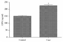
          The aim of this study was to investigate whether osteopontin (OPN) variants are associated with susceptibility to ankylosing spondylitis (AS) in a Chinese population. Polymorphisms at the 9175th position in exon 7 of OPN and rs17524488 were genotyped using direct sequencing in 186 unrelated AS patients and 188 ethnically matched healthy controls. Serum concentration of OPN was measured by enzyme-linked immunosorbent assay (ELISA) in all participants. AS patients displayed significantly higher OPN serum levels than the controls ( P < 001). A heterozygous, novel 9175 T>A in exon 7 of the OPN gene was found in this study. In healthy controls, subjects carrying the rs17524488 G/G genotype of the OPN display significantly higher OPN serum levels than the GG/GG genotype ( P < 0.05). Plasma OPN level is implicated as an early diagnostic marker of AS. The novel 9175th- (exon 7) position polymorphism of OPN and rs17524488 were related to susceptibility to AS in a Chinese population, the rs17524488 G/G genotype may be involved in the pathogenesis of AS, and the precise molecular mechanism underlying the influence of OPN polymorphisms on the development of AS remains to be determined in the further prospective studies.

          Related collections

          Most cited references26

          • Record: found
          • Abstract: found
          • Article: not found

          Osteopontin: role in immune regulation and stress responses.

          Recent research has led to a better but as yet incomplete understanding of the complex roles osteopontin plays in mammalian physiology. A soluble protein found in all body fluids, it stimulates signal transduction pathways (via integrins and CD44 variants) similar to those stimulated by components of the extracellular matrix. This appears to promote the survival of cells exposed to potentially lethal insults such as ischemia/reperfusion or physical/chemical trauma. OPN is chemotactic for many cell types including macrophages, dendritic cells, and T cells; it enhances B lymphocyte immunoglobulin production and proliferation. In inflammatory situations it stimulates both pro- and anti-inflammatory processes, which on balance can be either beneficial or harmful depending on what other inputs the cell is receiving. OPN influences cell-mediated immunity and has been shown to have Th1-cytokine functions. OPN deficiency is linked to a reduced Th1 immune response in infectious diseases, autoimmunity and delayed type hypersensitivity. OPN's role in the central nervous system and in stress responses has also emerged as an important aspect related to its cytoprotective and immune functions. Evidence suggests that either OPN or anti-OPN monoclonal antibodies (depending on the circumstances) might be clinically useful in modulating OPN function. Manipulation of plasma OPN levels may be useful in the treatment of autoimmune disease, cancer metastasis, osteoporosis and some forms of stress.
            Bookmark
            • Record: found
            • Abstract: found
            • Article: not found

            Expression, roles, receptors, and regulation of osteopontin in the kidney.

            Osteopontin (OPN) is a secreted glycoprotein in both phosphorylated and non-phosphorylated forms. It contains an Arg-Gly-Asp cell-binding sequence and a thrombin-cleavage site. OPN is mainly present in the loop of Henle and distal nephrons in normal kidneys in animals and humans. After renal damage, OPN expression may be significantly up-regulated in all tubule segments and glomeruli. Studies utilizing OPN gene-deficient mice, antisense-treated or anti-OPN-treated animals have demonstrated that OPN promotes accumulation of macrophages, and may play a role in macrophage-mediated renal injury, but that the effect may be mild and short-lived. On the other hand, OPN has some renoprotective actions in renal injury, such as increasing tolerance to acute ischemia, inhibiting inducible nitric oxide synthase and suppressing nitric oxide synthesis, reducing cell peroxide levels and promoting the survival of cells exposed to hypoxia, decreasing cell apoptosis and participating in the regeneration of cells. In addition, OPN is associated with renal stones, but whether it acts as a promoter or inhibitor of stone formation is controversial. It has been demonstrated that OPN receptors include two families: integrin and CD44. The OPN integrin receptors include alpha(v)beta(3), alpha(v)beta(1), alpha(v)beta(5) and alpha(9)beta(1), and alpha(4)beta(1). In normal human kidneys, standard CD44 is expressed most dominantly. Different OPN functions are mediated via distinct receptors. Parathyroid hormone, vitamin D(3), calcium, phosphate and some cytokines increase OPN expression in vitro or in vivo, whereas female sex hormones and angiotensin-converting enzyme inhibitors or angiotensin II receptor antagonists decrease OPN expression in some renal damage states.
              Bookmark
              • Record: found
              • Abstract: found
              • Article: not found

              Polymorphisms in the osteopontin promoter affect its transcriptional activity.

              Understanding the molecular mechanisms that underlie regulation of transcription of the human osteopontin encoding gene (OPN) may help to clarify several processes, such as fibrotic evolution of organ damage, tumorigenesis and metastasis, and immune response, in which OPN overexpression is observed. With the aim to evaluate variants with functional effect on transcription, we have analyzed the promoter region and focused our investigation on three common variants present in the first 500 bp upstream of the transcription start site. Transfection of constructs carrying the four most frequent haplotypes relative to variants at -66, -156, and -443 fused to the luciferase reporter gene in a panel of different cell lines showed that one haplotype conferred a significantly reduced level of reporter gene expression in all tested cell lines. We describe that the -66 polymorphism modifies the binding affinity for the SP1/SP3 transcription factors, the -156 polymorphism is included in a yet uncharacterized RUNX2 binding site, and the -443 polymorphism causes differential binding of an unknown factor. The finding of differential effects of various combination of variants in haplotypes may contribute to explain data of association studies reported in several already published articles. Future association studies using haplotypes instead of single OPN variants will allow to achieve more accurate results referable to differential expression of OPN in several common diseases, in which OPN is considered a candidate susceptibility gene.
                Bookmark

                Author and article information

                Contributors
                Journal
                Biomed Res Int
                Biomed Res Int
                BMRI
                BioMed Research International
                Hindawi
                2314-6133
                2314-6141
                2018
                18 January 2018
                : 2018
                : 3458439
                Affiliations
                1Department of Pharmacy, The Central Hospital of Wuhan, Tongji Medical College, Huazhong University of Science and Technology, Wuhan, Hubei, China
                2Department of Pain, The Central Hospital of Wuhan, Tongji Medical College, Huazhong University of Science and Technology, Wuhan, Hubei, China
                3Department of Endocrinology, The Central Hospital of Wuhan, Tongji Medical College, Huazhong University of Science and Technology, Wuhan, Hubei, China
                4Department of Information, The Central Hospital of Wuhan, Tongji Medical College, Huazhong University of Science and Technology, Wuhan, Hubei, China
                Author notes

                Academic Editor: Ewa Mojs

                Author information
                http://orcid.org/0000-0003-2119-7570
                http://orcid.org/0000-0002-7576-2945
                Article
                10.1155/2018/3458439
                5822803
                64f57274-1d0a-4691-89d1-7bb1057d2012
                Copyright © 2018 Juyi Li et al.

                This is an open access article distributed under the Creative Commons Attribution License, which permits unrestricted use, distribution, and reproduction in any medium, provided the original work is properly cited.

                History
                : 26 October 2017
                : 20 December 2017
                Funding
                Funded by: Health and Family Planning Commission of Wuhan Municipality
                Award ID: WX17B02
                Funded by: Health and Family Planning Commission of Hubei Province
                Award ID: WJ2017M183
                Categories
                Research Article

                Comments

                Comment on this article