3
views
0
recommends
+1 Recommend
2 collections
    0
    shares
      • Record: found
      • Abstract: found
      • Article: found
      Is Open Access

      Nitric oxide signaling in ctenophores

      research-article

      Read this article at

      Bookmark
          There is no author summary for this article yet. Authors can add summaries to their articles on ScienceOpen to make them more accessible to a non-specialist audience.

          Abstract

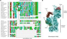
          Nitric oxide (NO) is one of the most ancient and versatile signal molecules across all domains of life. NO signaling might also play an essential role in the origin of animal organization. Yet, practically nothing is known about the distribution and functions of NO-dependent signaling pathways in representatives of early branching metazoans such as Ctenophora. Here, we explore the presence and organization of NO signaling components using Mnemiopsis and kin as essential reference species. We show that NO synthase (NOS) is present in at least eight ctenophore species, including Euplokamis and Coeloplana, representing the most basal ctenophore lineages. However, NOS could be secondarily lost in many other ctenophores, including Pleurobrachia and Beroe. In Mnemiopsis leidyi, NOS is present both in adult tissues and differentially expressed in later embryonic stages suggesting the involvement of NO in developmental mechanisms. Ctenophores also possess soluble guanylyl cyclases as potential NO receptors with weak but differential expression across tissues. Combined, these data indicate that the canonical NO-cGMP signaling pathways existed in the common ancestor of animals and could be involved in the control of morphogenesis, cilia activities, feeding and different behaviors.

          Related collections

          Most cited references97

          • Record: found
          • Abstract: found
          • Article: found
          Is Open Access

          Highly accurate protein structure prediction with AlphaFold

          Proteins are essential to life, and understanding their structure can facilitate a mechanistic understanding of their function. Through an enormous experimental effort 1 – 4 , the structures of around 100,000 unique proteins have been determined 5 , but this represents a small fraction of the billions of known protein sequences 6 , 7 . Structural coverage is bottlenecked by the months to years of painstaking effort required to determine a single protein structure. Accurate computational approaches are needed to address this gap and to enable large-scale structural bioinformatics. Predicting the three-dimensional structure that a protein will adopt based solely on its amino acid sequence—the structure prediction component of the ‘protein folding problem’ 8 —has been an important open research problem for more than 50 years 9 . Despite recent progress 10 – 14 , existing methods fall far short of atomic accuracy, especially when no homologous structure is available. Here we provide the first computational method that can regularly predict protein structures with atomic accuracy even in cases in which no similar structure is known. We validated an entirely redesigned version of our neural network-based model, AlphaFold, in the challenging 14th Critical Assessment of protein Structure Prediction (CASP14) 15 , demonstrating accuracy competitive with experimental structures in a majority of cases and greatly outperforming other methods. Underpinning the latest version of AlphaFold is a novel machine learning approach that incorporates physical and biological knowledge about protein structure, leveraging multi-sequence alignments, into the design of the deep learning algorithm. AlphaFold predicts protein structures with an accuracy competitive with experimental structures in the majority of cases using a novel deep learning architecture.
            Bookmark
            • Record: found
            • Abstract: found
            • Article: found
            Is Open Access

            SWISS-MODEL: homology modelling of protein structures and complexes

            Abstract Homology modelling has matured into an important technique in structural biology, significantly contributing to narrowing the gap between known protein sequences and experimentally determined structures. Fully automated workflows and servers simplify and streamline the homology modelling process, also allowing users without a specific computational expertise to generate reliable protein models and have easy access to modelling results, their visualization and interpretation. Here, we present an update to the SWISS-MODEL server, which pioneered the field of automated modelling 25 years ago and been continuously further developed. Recently, its functionality has been extended to the modelling of homo- and heteromeric complexes. Starting from the amino acid sequences of the interacting proteins, both the stoichiometry and the overall structure of the complex are inferred by homology modelling. Other major improvements include the implementation of a new modelling engine, ProMod3 and the introduction a new local model quality estimation method, QMEANDisCo. SWISS-MODEL is freely available at https://swissmodel.expasy.org.
              Bookmark
              • Record: found
              • Abstract: found
              • Article: not found

              The Phyre2 web portal for protein modeling, prediction and analysis.

              Phyre2 is a suite of tools available on the web to predict and analyze protein structure, function and mutations. The focus of Phyre2 is to provide biologists with a simple and intuitive interface to state-of-the-art protein bioinformatics tools. Phyre2 replaces Phyre, the original version of the server for which we previously published a paper in Nature Protocols. In this updated protocol, we describe Phyre2, which uses advanced remote homology detection methods to build 3D models, predict ligand binding sites and analyze the effect of amino acid variants (e.g., nonsynonymous SNPs (nsSNPs)) for a user's protein sequence. Users are guided through results by a simple interface at a level of detail they determine. This protocol will guide users from submitting a protein sequence to interpreting the secondary and tertiary structure of their models, their domain composition and model quality. A range of additional available tools is described to find a protein structure in a genome, to submit large number of sequences at once and to automatically run weekly searches for proteins that are difficult to model. The server is available at http://www.sbg.bio.ic.ac.uk/phyre2. A typical structure prediction will be returned between 30 min and 2 h after submission.
                Bookmark

                Author and article information

                Contributors
                Journal
                Front Neurosci
                Front Neurosci
                Front. Neurosci.
                Frontiers in Neuroscience
                Frontiers Media S.A.
                1662-4548
                1662-453X
                22 March 2023
                2023
                : 17
                : 1125433
                Affiliations
                [1] 1Department of Neuroscience, McKnight Brain Institute, University of Florida , Gainesville, FL, United States
                [2] 2The Whitney Laboratory for Marine Bioscience, University of Florida , St. Augustine, FL, United States
                [3] 3Institute of Higher Nervous Activity and Neurophysiology of RAS , Moscow, Russia
                Author notes

                Edited by: James Newcomb, New England College, United States

                Reviewed by: Thibaut Brunet, Pasteur Institute, France; Josean Reyes-Rivera, Pasteur Institute, France, in collaboration with reviewer TB; Rhanor Gillette, University of Illinois Urbana-Champaign, United States

                This article was submitted to Neural Technology, a section of the journal Frontiers in Neuroscience

                Article
                10.3389/fnins.2023.1125433
                10073611
                37034176
                6519103f-af74-4e9d-aa1a-d4e3cd4d5d50
                Copyright © 2023 Moroz, Mukherjee and Romanova.

                This is an open-access article distributed under the terms of the Creative Commons Attribution License (CC BY). The use, distribution or reproduction in other forums is permitted, provided the original author(s) and the copyright owner(s) are credited and that the original publication in this journal is cited, in accordance with accepted academic practice. No use, distribution or reproduction is permitted which does not comply with these terms.

                History
                : 16 December 2022
                : 27 February 2023
                Page count
                Figures: 11, Tables: 0, Equations: 0, References: 97, Pages: 15, Words: 9067
                Funding
                Funded by: National Institute of Neurological Disorders and Stroke, doi 10.13039/100000065;
                This work was supported in part by the Human Frontiers Science Program (RGP0060/2017) and the National Science Foundation (1146575, 1557923, 1548121, and 1645219) grants to LM. Research reported in this publication was also supported in part by the National Institute of Neurological Disorders and Stroke of the National Institutes of Health under Award Number R01NS114491 (to LM).
                Categories
                Neuroscience
                Original Research

                Neurosciences
                mnemiopsis,ctenophora,nitric oxide synthase,guanylate cyclase,pleurobrachia,nervous system evolution,porifera,placozoa

                Comments

                Comment on this article