7
views
0
recommends
+1 Recommend
0 collections
    0
    shares
      • Record: found
      • Abstract: found
      • Article: found
      Is Open Access

      A huge haemorrhagic suprarenal pseudocyst: an unusual presentation of a rare condition

      case-report
      1 , 2 , , 1
      BMJ Case Reports
      BMJ Publishing Group
      general practice / family medicine, adrenal disorders

      Read this article at

      Bookmark
          There is no author summary for this article yet. Authors can add summaries to their articles on ScienceOpen to make them more accessible to a non-specialist audience.

          Abstract

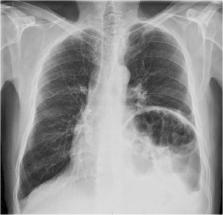
          Haemorrhagic suprarenal pseudocysts are very rare and are often incidental findings at surgery or autopsy, though they can sometimes present with predominantly gastrointestinal or endocrine symptoms, including intraperitoneal bleeding or sepsis. Our case report is of a 48-year-old man who presented at our primary healthcare centre with 2-month history of predominantly respiratory symptoms of cough and shortness of breath. CT scan revealed a suprarenal cyst measuring 14.2×13.5×13.1 cm for which he was operated and made a full recovery. A detailed literature review reveals that there has never been a case of a haemorrhagic suprarenal pseudocyst presenting with predominantly respiratory symptoms, which is why we decided to document this case report.

          Related collections

          Most cited references9

          • Record: found
          • Abstract: found
          • Article: not found

          Cystic lesions of the adrenal gland: our experience over the last 20 years.

          Cystic lesions of the adrenal gland are uncommon, often presenting with nonspecific clinical and radiologic findings, and are thus underrecognized. They are occasionally associated with malignant neoplasms, which can greatly mimic benign lesions and carry detrimental clinical consequences if misdiagnosed. Here we present our 20-year experience (1992-2012) with these lesions at an academic medical center. Among more than 4500 adrenal gland specimens, 31 cases of adrenal lesions with a predominant cystic component were identified in 30 patients with an age range of 34 to 86 years (median, 55.5 years) and a male/female ratio of 13:17. Macroscopic descriptions, available histologic and immunostain slides, and available radiologic records were reviewed for all included cases. Radiologic studies and gross examination correlated well, and hemorrhage (26 cases; 84%) and encapsulation (25 cases; 81%) appeared to be nonspecific radiologic/gross features shared across histologic subtypes. Microscopic review identified 12 cases (39%) of pseudocysts, 2 cases (6%) of endothelium-derived cysts, and 17 cases (55%) of epithelium-derived cysts. Among these 31 cystic adrenal lesions, 2 cases (6%) were malignant neoplasms (1 epithelioid angiosarcoma, 1 adrenocortical carcinoma). Radiologic impression and histopathologic diagnosis were concordant in 11 (73%) of the 15 cases for which radiologic records were available. This study represents the second largest case series to date on cystic adrenal lesions and presents a comprehensive review on their demographic, clinical, radiologic, and gross and microscopic pathologic features, as well as their differential diagnoses.
            Bookmark
            • Record: found
            • Abstract: found
            • Article: not found

            Adrenal pseudocyst: a clinical and pathologic study of eight cases.

            Adrenal pseudocysts are cystic lesions arising within the adrenal gland surrounded by a fibrous tissue wall devoid of a recognizable lining layer. This study comprised eight adrenal pseudocysts surgically excised at the Massachusetts General Hospital. The median age of the patients was 41 years. Seven of the eight individuals were women. There was no apparent etiologic relationship to prior trauma or pregnancy. Half of the patients described symptoms that resolved following pseudocyst removal. The remaining individuals were asymptomatic with adrenal pseudocysts discovered incidentally during the work up of other medical problems. Adrenal pseudocyst size ranged from 1.8 to 10 cm. Pseudocyst size did not correlate with the presence of symptoms. The histologic and immunohistochemical findings in this study suggest that at least some adrenal pseudocysts are of vascular origin. In two lesions, small foci of residual cells lining the inner pseudocyst wall were found that expressed factor VIII antigen (also Ulex Europaeus lectin in one case) but not vimentin, keratin, or epithelial membrane antigen. In both cases, abundant elastic tissue was present in the pseudocyst wall, and in one lesion, adrenal vein smooth muscle was present as well. In both cases, dilated sinusoids were found at the periphery of the pseudocysts and, in one lesion, the sinusoids appeared to coalesce to form the pseudocyst cavity. In the majority of the remaining six cases there was also some histologic evidence to suggest a vascular origin. In five and three cases, respectively, abundant elastic tissue and adrenal vein smooth muscle were found within the pseudocyst wall. In two lesions, both elastic tissue and smooth muscle were present. In addition, adrenal sinusoids were prominently dilated at the periphery of four pseudocysts and, in one case, the sinusoids appeared to coalesce to form the pseudocyst cavity.
              Bookmark
              • Record: found
              • Abstract: not found
              • Article: not found
              Is Open Access

              Adrenal pseudocyst: Diagnosis and laparoscopic management – A case report

                Bookmark

                Author and article information

                Journal
                BMJ Case Rep
                BMJ Case Rep
                bmjcr
                bmjcasereports
                BMJ Case Reports
                BMJ Publishing Group (BMA House, Tavistock Square, London, WC1H 9JR )
                1757-790X
                2021
                4 February 2021
                4 February 2021
                : 14
                : 2
                : e235158
                Affiliations
                [1 ]departmentDepartment of Family Medicine, Al Thumama Health Centre , Primary Health Care Corporation , Doha, Qatar
                [2 ]departmentDepartment of Family Medicine , Weill Cornell Medicine , Doha, Qatar
                Author notes
                [Correspondence to ] Dr Adekunle A Olowu; kunletinu@ 123456hotmail.com
                Author information
                http://orcid.org/0000-0002-6231-318X
                Article
                bcr-2020-235158
                10.1136/bcr-2020-235158
                7868216
                33542027
                6683e5aa-9ec4-4854-85df-6942d2752b2f
                © BMJ Publishing Group Limited 2021. Re-use permitted under CC BY-NC. No commercial re-use. See rights and permissions. Published by BMJ.

                This is an open access article distributed in accordance with the Creative Commons Attribution Non Commercial (CC BY-NC 4.0) license, which permits others to distribute, remix, adapt, build upon this work non-commercially, and license their derivative works on different terms, provided the original work is properly cited and the use is non-commercial. See:  http://creativecommons.org/licenses/by-nc/4.0/.

                History
                : 10 January 2021
                Funding
                Funded by: Primary Health Care Corporation Qatar;
                Award ID: PHCC/DCR/2020/04/037
                Categories
                Case Report
                1506
                157
                1325
                75
                Custom metadata
                unlocked

                general practice / family medicine,adrenal disorders

                Comments

                Comment on this article