28
views
0
recommends
+1 Recommend
0 collections
    0
    shares
      • Record: found
      • Abstract: found
      • Article: found
      Is Open Access

      Slc26a7 Chloride Channel Activity and Localization in Mouse Reissner’s Membrane Epithelium

      research-article

      Read this article at

      Bookmark
          There is no author summary for this article yet. Authors can add summaries to their articles on ScienceOpen to make them more accessible to a non-specialist audience.

          Abstract

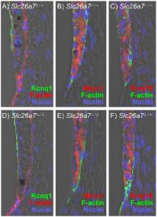
          Several members of the SLC26 gene family have highly-restricted expression patterns in the auditory and vestibular periphery and mutations in mice of at least two of these (SLC26A4 and SLC26A5) lead to deficits in hearing and/or balance. A previous report pointed to SLC26A7 as a candidate gene important for cochlear function. In the present study, inner ears were assayed by immunostaining for Slc26a7 in neonatal and adult mice. Slc26a7 was detected in the basolateral membrane of Reissner’s membrane epithelial cells but not neighboring cells, with an onset of expression at P5; gene knockout resulted in the absence of protein expression in Reissner’s membrane. Whole-cell patch clamp recordings revealed anion currents and conductances that were elevated for NO 3 over Cl and inhibited by I and NPPB. Elevated NO 3 currents were absent in Slc26a7 knockout mice. There were, however, no major changes to hearing (auditory brainstem response) of knockout mice during early adult life under constitutive and noise exposure conditions. The lack of Slc26a7 protein expression found in the wild-type vestibular labyrinth was consistent with the observation of normal balance. We conclude that SLC26A7 participates in Cl transport in Reissner’s membrane epithelial cells, but that either other anion pathways, such as ClC-2, possibly substitute satisfactorily under the conditions tested or that Cl conductance in these cells is not critical to cochlear function. The involvement of SLC26A7 in cellular pH regulation in other epithelial cells leaves open the possibility that SLC26A7 is needed in Reissner’s membrane cells during local perturbations of pH.

          Related collections

          Most cited references28

          • Record: found
          • Abstract: found
          • Article: not found

          Prestin is required for electromotility of the outer hair cell and for the cochlear amplifier.

          Hearing sensitivity in mammals is enhanced by more than 40 dB (that is, 100-fold) by mechanical amplification thought to be generated by one class of cochlear sensory cells, the outer hair cells. In addition to the mechano-electrical transduction required for auditory sensation, mammalian outer hair cells also perform electromechanical transduction, whereby transmembrane voltage drives cellular length changes at audio frequencies in vitro. This electromotility is thought to arise through voltage-gated conformational changes in a membrane protein, and prestin has been proposed as this molecular motor. Here we show that targeted deletion of prestin in mice results in loss of outer hair cell electromotility in vitro and a 40-60 dB loss of cochlear sensitivity in vivo, without disruption of mechano-electrical transduction in outer hair cells. In heterozygotes, electromotility is halved and there is a twofold (about 6 dB) increase in cochlear thresholds. These results suggest that prestin is indeed the motor protein, that there is a simple and direct coupling between electromotility and cochlear amplification, and that there is no need to invoke additional active processes to explain cochlear sensitivity in the mammalian ear.
            Bookmark
            • Record: found
            • Abstract: found
            • Article: not found

            A physiological place-frequency map of the cochlea in the CBA/J mouse.

            Genetically manipulated mice have gained a prominent role in in vivo research on development and function of the auditory system. A prerequisite for the interpretation of normal and abnormal structural and functional features of the inner ear is the exact knowledge of the cochlear place-frequency map. Using a stereotaxic approach to the projection site of the auditory nerve fibers in the cochlear nucleus, we succeeded in labelling physiologically characterized auditory nerve afferents and determined their peripheral innervation site in the cochlea. From the neuronal characteristic frequency (CF) and the innervation site in the organ of Corti a place-frequency map was established for characteristic frequencies between 7.2 and 61.8 kHz, corresponding to locations between 90% and 10% basilar membrane length (base = 0%, apex = 100%, mean length measured under the inner hair cells 5.13 mm). The relation between normalized distance from the base (d) and frequency (kHz) can be described by a simple logarithmic function: d(%) = 156.5-82.5 x log(f), with a slope of 1.25 mm/octave of frequency. The present map, recorded under physiological conditions, differs from earlier maps determined with different methods. The simple logarithmic place-frequency relation found in the mouse indicates that mice are acoustic generalists rather than specialists.
              Bookmark
              • Record: found
              • Abstract: found
              • Article: not found

              Gap junctions in the rat cochlea: immunohistochemical and ultrastructural analysis.

              Gap junctions in the rat cochlea were investigated using immunostaining for connexin26 and transmission electron microscopy. Electron microscopy of normal and pre-embedded immunostained material showed that there were gap junctions between and among all cells that light microscopy showed to have immunostained appositions. Light microscopy showed immunostaining between and among cell types that electron microscopy showed to be joined by gap junctions. Immunostaining for connexin26 was therefore taken as providing a reasonable approximation of the locations of gap junctions throughout the cochlea and was used to provide an overview of the extent of those locations. Cells interconnected via gap junctions fell into one of two groups. The first group consists of nonsensory epithelial cells and includes interdental cells of the spiral limbus, inner sulcus cells, organ of Corti supporting cells, outer sulcus cells, and cells within the root processes of the spiral ligament. The second group consists of connective tissue cells and includes various fibrocyte types of the spiral limbus and spiral ligament, basal and intermediate cells of the stria vascularis, and mesenchymal cells which line the scala vestibuli. The present work represents a first attempt towards a description of how serial gap junctions among cochlear cells reflect a level of organization of the tissue. The organization described here, together with a great deal of information from previous investigators, suggest that serially arranged gap junctions of both epithelial and connective tissue cells serve as the structural basis for recycling endolymphatic potassium ions that pass through the sensory cells during the transduction process.
                Bookmark

                Author and article information

                Contributors
                Role: Editor
                Journal
                PLoS One
                PLoS ONE
                plos
                plosone
                PLoS ONE
                Public Library of Science (San Francisco, USA )
                1932-6203
                2014
                8 May 2014
                : 9
                : 5
                : e97191
                Affiliations
                [1 ]Anatomy & Physiology Department, Cellular Biophysics Laboratory, Kansas State University, Manhattan, Kansas, United States of America
                [2 ]Anatomy & Physiology Department, Cell Physiology Laboratory, Kansas State University, Manhattan, Kansas, United States of America
                [3 ]Department of Medicine and Center on Genetics of Transport, University of Cincinnati, Cincinnati, Ohio, United States of America
                Albany Medical College, United States of America
                Author notes

                Competing Interests: The authors have declared that no competing interests exist.

                Conceived and designed the experiments: KXK DCM PW. Performed the experiments: KXK JDS HMK DGH JX. Analyzed the data: KXK JDS DGH DCM PW. Contributed reagents/materials/analysis tools: DCM PW MS. Wrote the paper: KXK DCM PW.

                Article
                PONE-D-14-03406
                10.1371/journal.pone.0097191
                4014619
                24810589
                66ebb5a5-cf66-4ca8-a818-0c7ae7d77422
                Copyright @ 2014

                This is an open-access article distributed under the terms of the Creative Commons Attribution License, which permits unrestricted use, distribution, and reproduction in any medium, provided the original author and source are credited.

                History
                : 22 January 2014
                : 16 April 2014
                Page count
                Pages: 10
                Funding
                This work was supported by grants from the National Institutes of Health: R01-DC012151 (PW), R01-DC00212 (DCM), and P20-RR017686 (DCM). Further support (MS) was from a Merit Review award from the Department of Veterans Affairs and from the Center on Genetics of Transport and Epithelial Biology at the University of Cincinnati. The funders had no role in study design, data collection and analysis, decision to publish, or preparation of the manuscript.
                Categories
                Research Article
                Biology and Life Sciences
                Anatomy
                Body Fluids
                Fluid Physiology
                Cell Biology
                Cell Physiology
                Physiology
                Electrophysiology
                Neuroscience
                Sensory Systems
                Auditory System

                Uncategorized
                Uncategorized

                Comments

                Comment on this article