30
views
0
recommends
+1 Recommend
0 collections
    0
    shares
      • Record: found
      • Abstract: found
      • Article: found
      Is Open Access

      Isolated cysticercosis of the cauda equina

      case-report

      Read this article at

      Bookmark
          There is no author summary for this article yet. Authors can add summaries to their articles on ScienceOpen to make them more accessible to a non-specialist audience.

          Abstract

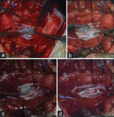
          Cysticercosis is the most common parasitic infection of the central nervous system. It is an endemic condition in developing countries, but the incidence rate is increasing in developed countries as well because of rising immigration. Spinal involvement is quite rare and it is usually associated with concomitant intracranial infective lesions. We present an unusual case of a 44-year-old woman who experienced a cauda equina syndrome. Magnetic resonance imaging disclosed two intradural cystic lesions at L4-L5 level. Only after histological examination the diagnosis of cysticercosis was definitively determined. The entire neuraxis evaluation confirmed that it was a rare form of isolated intradural racemosus type cysticercosis of the cauda equina. Steroids and albendazole were administered and post-operative course was uneventful. In this paper we discuss clinical, pathogenic and therapeutic aspects of this infective pathology.

          Related collections

          Most cited references10

          • Record: found
          • Abstract: found
          • Article: not found

          Isolated intradural-extramedullary spinal cysticercosis: a case report.

          Spinal cysticercosis is an uncommon manifestation of neurocysticercosis (NCC). We present a case of isolated lumbar intradural-extramedullary NCC. The patient was treated successfully with the surgical removal of the cyst. Spinal NCC should be considered in the differential diagnosis in high-risk populations with new symptoms suggestive of a spinal mass lesion. © 2011 International Society of Travel Medicine.
            Bookmark
            • Record: found
            • Abstract: found
            • Article: not found

            Medical and surgical treatment in neurocysticercosis a magnetic resonance study of 161 cases.

            In a prospective non-controlled study we have treated 161 consecutive cases of Active Neurocysticercosis (NCC) diagnosed by Magnetic Resonance (MR). Active NCC was classified in: (1) brain parenchymal cysts (85 cases); (2) ventricular cysts (24 cases); (3) subarachnoid cysts (46 cases); and (4) cysticercus racemose (6 cases). All patients had MR follow up 1 month after treatment. Twenty five patients had MR with gadopentetate dimeglumine (Gd) contrast enhancement. Cine MR was performed in one patient. Medical treatment with albendazole (ABZ) or Praziquantel (PZQ) was applied in 136 cases. Drug efficacy, assessed by disappearance of the lesion on MR, was 92.5% with ABZ and 60% with PZQ. Thirty patients were treated by surgery. Five patients of group I were treated surgically due either to refractory seizures or persistent abnormalities on MR. Ventricular cysts were removed in 20 cases; 4 cases with cysticercus racemose and one with subarachnoid spinal cyst were also treated by surgery. Two patients with 4th ventricle cysts received ABZ and ventricular shunt only. Two cases with intraventricular cysts (lateral ventricles) and two with racemose cysts were successfully treated with ABZ. The Gd infusion showed enhancement in cysts with adjacent inflammatory reaction or edema and in cases with meningeal inflammation. Cine MR was useful in the differential diagnosis with congenital arachnoid cyst. We conclude that (1) MR is sensitive in the diagnosis of active NCC and may be useful in evaluating degenerative changes in the parasite; (2) ABZ is highly effective in the treatment of parenchymal and subarachnoidal NCC; (3) Parenchymal lesions which remain with abnormal appearance on MR (Degenerative cysticerci or gliosis) and refractory seizures should be treated by surgery; (4) Cysticercus racemose without intracranial hypertension may be treated with ABZ; (5) Ventricular cysts are treated by surgical removal, however, ABZ and ventricular peritoneal shunt may also be an alternative approach.
              Bookmark
              • Record: found
              • Abstract: found
              • Article: not found

              Recurrent primary spinal subarachnoid neurocysticercosis.

              Case description. To describe a patient with a recurrent primary spinal subarachnoid neurocysticercosis (NCC) that was successfully treated with surgical decompression and medical therapy at our center. Spinal subarachnoid NCC is thought to be the secondary result from larval migration through the ventricular system into the spinal subarachnoid space. However, this entity can develop as a primary infection through blood stream or direct larval migration. It can result in high recurrence and severe neurologic morbidity if it is not treated in an appropriate manner. A 50-year-old woman with treatment history of spinal NCC presented with back pain and radicular pain. The lumbar magnetic resonance imaging showed a cystic lesion with septation and slight rim enhancement after gadolinium administration at the L4-S1 area. We performed surgical removal of this lesion and postoperative medical therapy for complete eradication of the parasite. The histopathology was diagnostic for a cysticercal cyst. Adjuvant medical therapy with albendazole was administered for 30 days after surgery. The patient remained symptom-free for 1 year after surgery without any evidence of recurrence. We report a rare case of recurrent primary spinal subarachnoid NCC at L4-S1 area. In cases of primary spinal subarachnoid NCC can be treated by adequate combined approach with surgery and medical therapy. Spinal subarachnoid NCC should be added to the differential diagnosis of primary spinal intradural cysts, because this lesion can occur primarily.
                Bookmark

                Author and article information

                Journal
                J Neurosci Rural Pract
                J Neurosci Rural Pract
                JNRP
                Journal of Neurosciences in Rural Practice
                Medknow Publications & Media Pvt Ltd (India )
                0976-3147
                0976-3155
                August 2013
                : 4
                : Suppl 1
                : S117-S119
                Affiliations
                [1] Department of Neurosurgery, Umberto I University General Hospital, Università Politecnica delle Marche, Ancona, Italy
                Author notes
                Address for correspondence: Dr. Maurizio Iacoangeli, Clinica di Neurochirurgia, Università Politecnica delle Marche, Ospedali Riuniti di Ancona, Via Conca #71, I – 60020, Torrette di Ancona, Italy. E-mail: neurotra@ 123456tiscali.it
                Article
                JNRP-4-117
                10.4103/0976-3147.116440
                3808039
                24174777
                67e57cdc-9507-4963-8458-abf1fb2fc32f
                Copyright: © Journal of Neurosciences in Rural Practice

                This is an open-access article distributed under the terms of the Creative Commons Attribution-Noncommercial-Share Alike 3.0 Unported, which permits unrestricted use, distribution, and reproduction in any medium, provided the original work is properly cited.

                History
                Categories
                Case Report

                Neurosciences
                cauda equina syndrome,isolated spinal cysticercosis,neurocysticercosis,parasitic infection,racemosus type,taenia solium

                Comments

                Comment on this article