- Record: found
- Abstract: found
- Article: found
Reconsidering the utility of manifest entrainment pacing in macroreentrant ventricular tachycardia: A case report
case-report
Masato Okada , MD,
Nobuaki Tanaka , MD,
Takafumi Oka , MD, PhD,
Koji Tanaka , MD,
Yuko Hirao , MD,
Koichi Inoue , MD, PhD
∗
03 June 2020
Read this article at
There is no author summary for this article yet. Authors can add summaries to their articles on ScienceOpen to make them more accessible to a non-specialist audience.
Abstract
Introduction
Entrainment pacing is a useful maneuver not only for establishing reentry as a mechanism
of tachycardia, but also for identifying a critical component of the circuit.
1
,
2
Concealed entrainment with postpacing interval (PPI) equal to the tachycardia cycle
length (TCL) strongly suggests the presence of the pacing site on the critical slow
conduction zone (SCZ) within the circuit.
1
,
2
On the other hand, manifest entrainment and orthodromic capture of the earliest activation
site (EAS) can demonstrate that the SCZ is located between the pacing site and the
EAS.
3
Manifest entrainment–guided catheter ablation targeting the “entrance” to the SCZ
is an effective method in the treatment of atrial tachycardia originating from the
vicinity of the atrioventricular node
4
and tricuspid annulus.
5
However, limited information is available regarding the effectiveness of the manifest
entrainment–guided strategy in the treatment of ventricular tachycardia (VT). We present
a case of macroreentrant VT that was successfully ablated by targeting the midmyocardial
“entrance” of critical SCZ identified using manifest entrainment pacing.
Case report
A 44-year-old man with unknown cardiomyopathy was referred to our hospital for an
electrical storm with repetitive appropriate implantable cardioverter-defibrillator
shocks. A hemodynamically stable and monomorphic wide QRS tachycardia on the 12-lead
electrocardiogram was incessantly observed in the emergency room (Supplemental Figure 1A).
The VT rate was 145 beats per minute (TCL = 412 ms), and the QRS morphology exhibited
a right bundle branch block configuration with left axis deviation, suggesting that
the location of the VT origin was inferior-posterior in the left ventricle (LV). The
physical examination, laboratory data, and echocardiographic data revealed no evidence
of acute coronary syndrome or myocarditis. The hypokinetic motion and thinning (5
mm) of the LV posterior wall remained unchanged with an LV ejection fraction of 48%.
Landilol and dexmedetomidine made the VT less frequent; however, treatment with medication
could not completely prevent the VT occurrence, and a long-term therapy-free survival
was strongly recommended, which made performing catheter ablation for the VT appropriate.
After informed consent was obtained, multipolar electrode catheters were inserted
into the femoral vein and positioned in the great cardiac vein and left ventricle
via a transseptal approach. Subsequently, heparin was administered to achieve an activated
clotting time of 250–300 seconds. The surface electrocardiogram and bipolar intracardiac
electrograms were continuously monitored and stored on a computer-based digital recording
system (LabSystem PRO; Bard Electrophysiology, Lowell, MA). The bipolar electrograms
were filtered from 30 to 500 Hz. Electroanatomic mapping was performed using the CARTO3
mapping system (Biosense Webster, Diamond Bar, CA). First, voltage mapping of the
LV was performed during sinus rhythm using a multielectrode PentaRay catheter (Biosense
Webster). LV scarring, defined as a bipolar electrogram amplitude of <1.5 mV, was
patchy, with a predilection for the basal inferior septum (Supplemental Figure 1B).
The endocardial unipolar voltage map showed additional remote low voltage area around
the inferior posterior of the LV (Supplement Figure 1C).
Pacing maneuvers were conducted using bipolar electrodes with fixed output (5 mA at
1.0 ms pulse width) generated by the stimulator (SEC-5104; Nihon Kohden, Tokyo, Japan).
Programmed ventricular stimulation at paced cycle lengths of 600 and 400 ms repeatedly
induced the clinical VT. Constant fusion and progressive fusion during the overdrive
pacing supported the tachycardia with a macroreentrant mechanism. Activation mapping
of the LV endomyocardium demonstrated a centrifugal spread with the endocardial EAS
(endo-EAS) located in the LV inferior septum. The total activation time within the
LV endocardium was 108 ms, much shorter than the TCL of 412 ms. Radiofrequency (RF)
energy in the range of 30–35 W was initially delivered to the endocardial EAS (endo-EAS)
during VT using an irrigated-tip catheter (ThermoCool ST SF; Biosense Webster). However,
the ablation of the endo-EAS did not modify the VT. Ventricular overdrive pacing at
that site exhibited manifest entrainment, a PPI − TCL (TCL subtracted from PPI) of
28 ms, and an electrograms-QRS equal to the paced QRS. These findings suggested that
the site was in an “outer loop” of the VT. Involvement of the extra-endocardium in
the reentrant circuit was suggested.
Before proceeding with the epicardial approach, we placed a microelectrode catheter
(EPstar 2F Fix microcatheter; Japan Lifeline, Tokyo, Japan) in the posterior lateral
vein to examine the epicardial potentials (Figure 1A). Discrete potentials (dull and
sharp potentials) were recorded on the electrocardiogram (Figure 1B). The dull and
sharp potentials were considered to be the far-field LV endocardial potential and
local epicardial potential, respectively. A sharp potential preceded the surface QRS
complex during the VT by 66 ms and became the EAS on the epicardium (epi-EAS). Concealed
entrainment, a PPI − TCL = 0 ms, and a stimulus-QRS = electrograms-QRS = 66 ms (16%
of TCL) identified the epi-EAS as the “exit” of the VT circuit (Figure 1C). From the
contralateral site, entrainment pacing with 5 mA at 1.0 ms pulse width was performed
using distal bipoles, which produced manifest entrainment and orthodromic capture
of the epi-EAS (Figure 2A). The result indicated that the “entrance” of the critical
SCZ was located within the ventricular myocardium between the pacing site and epi-EAS
(Figure 2B).
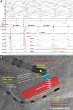
Figure 1
After insertion of a small electrode catheter into the posterior lateral vein and
placement of the MAP catheter on the endocardial surface opposite to the vein (A),
intracardiac electrograms were evaluated during both sinus rhythm and the ventricular
tachycardia (VT) (B). After 2 sinus beats, the VT was spontaneously initiated. The
sharp potential preceded the surface QRS complex by 66 ms during the VT, and the electrodes
of the EPI 1-2 became the earliest activation sites of the entire electrodes. Entrainment
pacing from the distal small electrodes (EPI 1-2) revealed entrainment with concealed
fusion (C). I, II, III, aVR, aVL, aVF, V1, V2, V3, V4, and V5 represent the surface
electrocardiogram leads; CS 1-2 to 19-20 represent the distal to proximal coronary
sinus recordings; MAP 1-2 represents the electrograms of the ablation catheter; and
EPI 1-2 to 7-8 represent the distal-to-proximal recordings of the small electrode
catheter. CS = coronary sinus; E-QRS = electrograms-QRS; PCL = pacing cycle length;
PPI = postpacing interval; S-QRS = stimulus-QRS; TCL = tachycardia cycle length.
Figure 2
A: Tracing during manifest entrainment by pacing delivered from the MAP catheter placed
on the opposite side of the epicardial earliest activation site (epi-EAS) is presented.
The asterisks indicate the electrograms captured by the last pacing stimulus, occurring
at the cycle length of 380 ms. The distal electrograms of the small microelectrode
catheter (EPI 1-2 and 2-3) are orthodromically captured with long conduction intervals
via the slow conduction zone (SCZ) (red asterisks), while the paroxysmal electrograms
are antidromically captured (green asterisks). The presence of the SCZ between the
pacing site and epi-EAS (orthodromic capture site) was suggested. B: Estimated ventricular
tachycardia (VT) circuit is shown with a white arrow. Given the limited mapping and
unknown captured area during entrainment pacing, it is also possible that the VT circuit
is completely midmyocardium/epicardium. CS = coronary sinus; PCL = pacing cycle length;
PPI = postpacing interval; RFCA = radiofrequency catheter ablation; TCL = tachycardia
cycle length.
RF energy applications to the epi-EAS were difficult because of the small diameter
of the vein and high tissue impedance. Therefore, we applied 30 W of RF energy from
the endocardium targeting the SCZ between the epi-EAS (exit) and contralateral endocardial
site (entrance side), which successfully terminated the VT in 7.5 seconds. Spatiotemporal
dynamics of contact (10–40 g) governed by cardiac and respiratory motion increased
the risk of the complications using high power. From the safety point of view, RF
energy within the range of 30–35 W was repeatedly delivered until the ablation index
reached 500 (estimated lesion depth = 5 mm equivalent to posterior wall thickness).
No abnormal low-amplitude and fractionated electrograms, suggestive of slow conduction,
were observed at the successful ablation site (Figure 3). The procedure was completed
without any complications after confirming the noninducibility of the clinical VT
even after an isoproterenol infusion. The patient was discharged 3 days after the
CA with continuation of amiodarone (100 mg/d). During the 2-year observation period
after the procedure, an appropriate implantable cardioverter-defibrillator shock for
VT was experienced once in 15 months; however, no further VT storms have been observed
following the ablation procedure at the time of writing this report.
Figure 3
A: The fluoroscopic positions and local electrograms were compared between the initial
radiofrequency catheter ablation (RFCA) site and successful RFCA site. Abnormal, low-amplitude,
fractionated electrograms preceding the QRS were observed at the initial RFCA site.
However, we did not record such abnormal potentials at the successful RFCA site. B:
The anatomical position projected by the CARTO activation map (Biosense Webster, Diamond
Bar, CA) showed that the successful RFCA site was inferior and lateral to the left
ventricle and 38 mm remote from the initial RFCA site. INF = inferior; LAO = left
anterior oblique; RAO = right anterior oblique; RFCA = radiofrequency catheter ablation.
Discussion
The role of entrainment in identifying the ablation target has become less important
with the evolution of 3-dimensional (3D) electroanatomic mapping systems. However,
increasing awareness of the limitations of a visual representation
6
and recognition of the 3D nature of VT circuits
7
remind us of the importance of entrainment pacing. Owing to this entrainment technique,
we can identify the critical components of macroreentrant tachycardias even without
the complete delineation of the circuit. In our case, involvement of the midmyocardium
in the reentrant circuit prevented the visualization of the entire circuit on the
endocardial 3D mapping system. However, we identified the “exit” and “entrance” of
the SCZ by using concealed and manifest entrainment.
Precisely speaking, the site with manifest entrainment was an “outer loop” and was
different from the “entrance” of the critical SCZ. However, in combination with orthodromic
capture of the EAS, we could identify the direction of the “entrance” from the pacing
site. For a more accurate determination of the proximal site of the “entrance,” mapping
during manifest entrainment is required to measure the time between the pacing and
the EAS electrogram.
3
The shorter the time, the more proximal is the pacing site to the “entrance.” Although
the anatomical opposite of the epi-EAS does not always mean the electrically shortest
endocardial site to the midmyocardial entrance, we applied RF energy from there because
of the relatively short distance from the endocardial ablation site to the epi-EAS
(5-mm wall thickness), which would enable the modification of the midmyocardial SCZ.
The other notable point in this case was that we could successfully approach the midmyocardial
SCZ from the seemingly healthy endocardium. Conventionally, the localization of the
SCZ in the midmyocardium is presumed by the discontinuity of the endocardial-epicardial
activation
7
; however, manifest entrainment and orthodromic capture of the EAS by pacing from
an opposite site of the EAS adds more direct electrophysiological evidence of a midmyocardial
SCZ.
Discussion about the captured area is required for the accurate interpretation of
entrainment pacing. Although changing pacing output can help differentiate between
far-field or near-field capture, we did not examine it in the present case. However,
the finding that PPI was equal to TCL (not PPI < TCL) at the successful ablation site
indicated that the paced activation wavefront returned on time and did not advance
by far-field downstream capture. This suggests that the captured area during entrainment
did not include the midmyocardial SCZ. Although it is difficult to conclude how deep
and how broad the endocardial pacing recruited myocardium, manifest entrainment and
orthodromic capture of EAS suggests that SCZ is located between the pacing site and
EAS.
Finally, although RF energy application from the epicardium is feasible, it is often
limited by epicardial fat and involves potential risks, such as pericardial bleeding
and collateral injury that includes the coronary vessels or phrenic nerve.
1
,
2
Moreover, repetitive access to the pericardial space potentially produces pericardial
adhesions, resulting in inaccessibility to the space for future treatment. A recent
study has shown that RF energy applications from the endocardium can modify the epicardial
substrate, especially in a region with wall thinning (<5 mm).
8
Therefore, an initial endocardial ablation at the critical site identified using entrainment
pacing would reduce the extent and in some cases the necessity of epicardial ablation.
Conclusions
This case highlighted the usefulness of entrainment pacing in the detection of a midmyocardial
isthmus of the VT circuit. Although the exact boundaries of the reentry circuit were
not convincingly defined, we elucidated the “exit” and “entrance” of the midmyocardial
SCZ using epicardial and endocardial entrainment pacing. Manifest entrainment–guided
catheter ablation strategy targeting the “entrance” of the SCZ can be an alternative
method for VT ablation.
Key Teaching Points
•
Entrainment pacing can help identify critical components of macroreentrant tachycardias
even without a complete delineation of the circuit.
•
Localization of the slow conduction zone (SCZ) in the midmyocardium would be more
convincingly elucidated with sandwiched entrainment from the endocardium and epicardium.
•
Manifest entrainment–guided catheter ablation strategy targeting the entrance of the
SCZ is an alternative method for ventricular tachycardia ablation.
Related collections
Most cited references7
- Record: found
- Abstract: found
- Article: not found
Exploring postinfarction reentrant ventricular tachycardia with entrainment mapping.
William G. Stevenson, Peter Friedman, Philip Sager … (1997)
- Record: found
- Abstract: found
- Article: not found
The Restructuring of Structural Heart Disease Practice During The Covid-19 Pandemic
Christine Chung, Tamim Nazif, Mariusz Wolbinski … (2020)
- Record: found
- Abstract: found
- Article: not found
Endocardial ablation to eliminate epicardial arrhythmia substrate in scar-related ventricular tachycardia.
Yuki Komatsu, Matthew Daly, Frédéric Sacher … (2014)