54
views
0
recommends
+1 Recommend
0 collections
    0
    shares
      • Record: found
      • Abstract: found
      • Article: found
      Is Open Access

      Grape seed proanthocyanidins inhibit the invasive potential of head and neck cutaneous squamous cell carcinoma cells by targeting EGFR expression and epithelial-to-mesenchymal transition

      research-article

      Read this article at

      Bookmark
          There is no author summary for this article yet. Authors can add summaries to their articles on ScienceOpen to make them more accessible to a non-specialist audience.

          Abstract

          Background

          Head and neck squamous cell carcinoma (HNSCC) is responsible for over 20,000 deaths every year in United States. Most of the deaths are due, in large part, to its propensity to metastasize. We have examined the effect of bioactive component grape seed proanthocyanidins (GSPs) on human cutaneous HNSCC cell invasion and the molecular mechanisms underlying these effects using SCC13 cell line as an in vitro model.

          Methods

          The therapeutic effects of GSPs on cancer cell invasion were studied using Boyden chamber and wound healing assays. The effects of GSPs on the levels of various proteins related with cancer cell invasion were determined using western blot analysis.

          Results

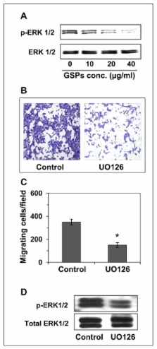
          Using in vitro cell invasion assays, we observed that treatment of SCC13 cells with GSPs resulted in a concentration-dependent inhibition of cell invasion of these cells, which was associated with a reduction in the levels of epidermal growth factor receptor (EGFR). Treatment of cells with gefitinib and erlotinib, inhibitors of EGFR, or transient transfection of SCC13 cells with EGFR small interfering RNA, also inhibited invasion of these cells. The inhibition of cell invasion by GSPs was associated with the inhibition of the phosphorylation of ERK1/2, a member of mitogen-activated protein kinase family. Treatment of cells with UO126, an inhibitor of MEK, also inhibited the invasion potential of SCC13 cells. Additionally, inhibition of human cutaneous HNSCC cell invasion by GSPs was associated with reversal of epithelial-to-mesenchymal transition (EMT) process, which resulted in an increase in the levels of epithelial biomarker (E-cadherin) while loss of mesenchymal biomarkers (vimentin, fibronectin and N-cadherin) in cells. Similar effect on EMT biomarkers was also observed when cells were treated with erlotinib.

          Conclusion

          The results obtained from this study indicate that grape seed proanthocyanidins have the ability to inhibit the invasion of human cutaneous HNSCC cells by targeting the EGFR expression and reversing the process of epithelial-to-mesenchymal transition. These data suggest that GSPs can be developed as a complementary and alternative medicine for the prevention of invasion/metastasis of HNSCC cells.

          Related collections

          Most cited references14

          • Record: found
          • Abstract: found
          • Article: not found

          Levels of TGF-alpha and EGFR protein in head and neck squamous cell carcinoma and patient survival.

          The most accurate predictor of disease recurrence in patients treated for head and neck squamous cell carcinoma is, at present, the extent of regional lymph node metastasis. Since elevated levels of epidermal growth factor receptor (EGFR) and of its ligand, transforming growth factor-alpha (TGF-alpha), have been detected in primary tumors of patients with head and neck squamous cell carcinoma, we determined whether tumor levels of these proteins were of prognostic importance. Monoclonal antibodies specific for EGFR and TGF-alpha were used for immunohistochemical detection of each protein in tissue sections of primary tumors from 91 patients who were treated by surgical resection. Levels of immunoreactive EGFR and TGF-alpha were quantified by use of a computerized image analysis system and were normalized to appropriate standards. The logrank test and proportional hazards regression analysis were used to calculate the probability that EGFR and TGF-alpha levels were associated with disease-free survival (i.e., no recurrence of cancer) and cause-specific survival (i.e., patients do not die of their disease). All P values were two-sided. When tumor levels of EGFR or TGF-alpha were analyzed as continuous variables, disease-free survival and cause-specific survival were reduced among patients with higher levels of EGFR (both P = .0001) or TGF-alpha (both P = .0001). In a multivariate analysis, tumor site, tumor level of EGFR, and tumor level of TGF-alpha were statistically significant predictors of disease-free survival; in a similar analysis, regional lymph node stage and tumor levels of EGFR and of TGF-alpha were significant predictors of cause-specific survival. Quantitation of EGFR and TGF-alpha protein levels in primary head and neck squamous cell carcinomas may be useful in identifying subgroups of patients at high risk of tumor recurrence and in guiding therapy.
            Bookmark
            • Record: found
            • Abstract: found
            • Article: not found

            Profiling early head and neck cancer.

            Head and neck squamous-cell carcinoma (HNSCC) is the sixth most common cancer worldwide and, disappointingly, survival rates are not improving. Moreover, HNSCC has a severe impact on the quality of life of patients and survivors, and the significant morbidity subsequent to treatment often mandates long-term multidisciplinary care, which places significant financial pressures on the treating institution. Therefore, prevention and early diagnosis of high-risk pre-malignant lesions are high priorities for reducing deaths due to head and neck cancer. Recent advances have begun to elucidate the different aetiologies of HNSCCs in relation to previous pre-malignancies and to identify which pre-malignant lesions are likely to progress to malignancy.
              Bookmark
              • Record: found
              • Abstract: found
              • Article: not found

              Quantitative immunohistochemical analysis of transforming growth factor-alpha and epidermal growth factor receptor in patients with squamous cell carcinoma of the head and neck.

              Transforming growth factor-alpha (TGF-alpha) and epidermal growth factor receptor (EGFR) mRNA are up-regulated in squamous cell carcinoma of the head and neck (SCCHN) tissues. Immunohistochemical staining with monoclonal antibodies to TGF-alpha and EGFR was undertaken to identify the cellular origin in tissue obtained from cancer patients and controls and to determine the correlation between mRNA expression levels and two methods of immunohistochemical evaluation. TGF-alpha protein staining occurred in the suprabasal layers and spared the basal layer of normal controls. Conversely, in histologically normal mucosa from SCCHN patients, TGF-alpha was present throughout the epithelium, including the basal layer. EGFR staining was negligible in normal mucosa from control patients without cancer and relatively increased in SCCHN tissues. Increasing staining intensity was correlated with worsening dysplasia and closer proximity to the tumor. Using computerized image analysis to quantify the intensity of immunostaining, the mean optical density (MOD) of TGF-alpha staining in histologically normal mucosa (P = 0.049) and tumors (P = 0.005) from SCCHN patients was significantly higher than in control normal mucosa from noncancer patients (1.9- and 1.7-fold, respectively). EGFR MOD was also greater in the histologically normal mucosa (P = 0.009) and tumors (P = 0.006) from SCCHN patients than in control normal mucosa (1.8- and 1.9-fold, respectively). For both TGF-alpha (P = 0.668) and EGFR (P = 0.116), the MOD was similar for both tumor and histologically normal mucosa from SCCHN patients. TGF-alpha and EGFR protein expression is increased early in head and neck squamous cell carcinogenesis and can be quantitated by computerized image analysis of immunohistochemical staining. Altered distribution of TGF-alpha protein in histologically normal mucosa from SCCHN patients compared with control mucosa from patients without cancer suggests a switch from a paracrine to an autocrine pathway.
                Bookmark

                Author and article information

                Journal
                BMC Complement Altern Med
                BMC Complementary and Alternative Medicine
                BioMed Central
                1472-6882
                2011
                21 December 2011
                : 11
                : 134
                Affiliations
                [1 ]Department of Dermatology, University of Alabama at Birmingham, Birmingham, AL, USA
                [2 ]Department of Surgery-Otolaryngology, University of Alabama at Birmingham, Birmingham, AL, USA
                [3 ]Comprehensive Cancer Center, University of Alabama at Birmingham, Birmingham, AL, USA
                [4 ]Nutrition Obesity Research Center, University of Alabama at Birmingham, Birmingham, AL, USA
                [5 ]Birmingham Veterans Affairs Medical Center, Birmingham, AL, 35294, USA
                Article
                1472-6882-11-134
                10.1186/1472-6882-11-134
                3258217
                22188922
                6b2419d3-9b89-4ff8-9d2d-19ff4c629fed
                Copyright ©2011 Sun et al; licensee BioMed Central Ltd.

                This is an Open Access article distributed under the terms of the Creative Commons Attribution License ( http://creativecommons.org/licenses/by/2.0), which permits unrestricted use, distribution, and reproduction in any medium, provided the original work is properly cited.

                History
                : 30 September 2011
                : 21 December 2011
                Categories
                Research Article

                Complementary & Alternative medicine
                Complementary & Alternative medicine

                Comments

                Comment on this article