52
views
0
recommends
+1 Recommend
0 collections
    0
    shares
      • Record: found
      • Abstract: found
      • Article: not found

      Competitive regulation of synaptic Ca influx by D2 dopamine and A2A adenosine receptors

      research-article
      ,
      Nature neuroscience

      Read this article at

      ScienceOpenPublisherPMC
      Bookmark
          There is no author summary for this article yet. Authors can add summaries to their articles on ScienceOpen to make them more accessible to a non-specialist audience.

          Abstract

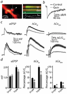
          Striatal D2-type dopamine receptors (D2Rs) are implicated in the pathophysiology of neuropsychiatric disorders, including Parkinson’s disease and schizophrenia. Although these receptors regulate striatal synaptic plasticity, the mechanisms underlying dopaminergic modulation of glutamatergic synapses are unclear. We combined optogenetics, 2-photon microscopy, and glutamate uncaging to examine D2R-dependent modulation of glutamatergic synaptic transmission in mouse striatopallidal neurons. We find that D2R activation reduces corticostriatal glutamate release and attenuates both synaptic- and action potential-evoked Ca influx into dendritic spines by approximately 50%. Modulation of Ca signaling is mediated by a PKA-dependent regulation of Ca entry through NMDA-type glutamate receptors that is inhibited by D2Rs and enhanced by activation of 2A-type adenosine receptors (A2ARs). D2Rs also produce a PKA- and A2AR-independent reduction in Ca influx through R-type voltage-gated Ca channels. These findings reveal that dopamine regulates spine Ca by multiple pathways and that competitive modulation of PKA controls NMDAR-mediated Ca signaling in the striatum.

          Related collections

          Most cited references38

          • Record: found
          • Abstract: found
          • Article: not found

          D1 and D2 dopamine-receptor modulation of striatal glutamatergic signaling in striatal medium spiny neurons.

          Dopamine shapes a wide variety of psychomotor functions. This is mainly accomplished by modulating cortical and thalamic glutamatergic signals impinging upon principal medium spiny neurons (MSNs) of the striatum. Several lines of evidence suggest that dopamine D1 receptor signaling enhances dendritic excitability and glutamatergic signaling in striatonigral MSNs, whereas D2 receptor signaling exerts the opposite effect in striatopallidal MSNs. The functional antagonism between these two major striatal dopamine receptors extends to the regulation of synaptic plasticity. Recent studies, using transgenic mice in which cells express D1 and D2 receptors, have uncovered unappreciated differences between MSNs that shape glutamatergic signaling and the influence of DA on synaptic plasticity. These studies have also shown that long-term alterations in dopamine signaling produce profound and cell-type-specific reshaping of corticostriatal connectivity and function.
            Bookmark
            • Record: found
            • Abstract: found
            • Article: not found

            Dopamine receptors: from structure to function.

            The diverse physiological actions of dopamine are mediated by at least five distinct G protein-coupled receptor subtypes. Two D1-like receptor subtypes (D1 and D5) couple to the G protein Gs and activate adenylyl cyclase. The other receptor subtypes belong to the D2-like subfamily (D2, D3, and D4) and are prototypic of G protein-coupled receptors that inhibit adenylyl cyclase and activate K+ channels. The genes for the D1 and D5 receptors are intronless, but pseudogenes of the D5 exist. The D2 and D3 receptors vary in certain tissues and species as a result of alternative splicing, and the human D4 receptor gene exhibits extensive polymorphic variation. In the central nervous system, dopamine receptors are widely expressed because they are involved in the control of locomotion, cognition, emotion, and affect as well as neuroendocrine secretion. In the periphery, dopamine receptors are present more prominently in kidney, vasculature, and pituitary, where they affect mainly sodium homeostasis, vascular tone, and hormone secretion. Numerous genetic linkage analysis studies have failed so far to reveal unequivocal evidence for the involvement of one of these receptors in the etiology of various central nervous system disorders. However, targeted deletion of several of these dopamine receptor genes in mice should provide valuable information about their physiological functions.
              Bookmark
              • Record: found
              • Abstract: found
              • Article: not found

              Cortical excitatory neurons and glia, but not GABAergic neurons, are produced in the Emx1-expressing lineage.

              By homologous recombination of an internal ribosome entry site and Cre recombinase coding region into the 3'-untranslated region of the mouse Emx1 gene, we have generated a strain of mice, Emx1(IRES)cre, that expresses the Cre recombinase in a spatial and temporal pattern like that observed for Emx1. When mated to reporter strains, these mice are a sensitive means to fate-map the Emx1-expressing cells of the developing forebrain. Our results demonstrate that radial glia, Cajal-Retzius cells, glutamatergic neurons, astrocytes, and oligodendrocytes of most pallial structures originate from an Emx1-expressing lineage. On the other hand, most of the pallial GABAergic neurons arise outside the Emx1-expressing lineage. Structures that are located near the basal ganglia (e.g., the amygdala and endopiriform nuclei) are not uniformly derived from Emx1-expressing cells.
                Bookmark

                Author and article information

                Journal
                9809671
                21092
                Nat Neurosci
                Nature neuroscience
                1097-6256
                1546-1726
                10 June 2010
                4 July 2010
                August 2010
                1 February 2011
                : 13
                : 8
                : 958-966
                Affiliations
                Howard Hughes Medical Institute, Department of Neurobiology, Harvard Medical School, Boston, MA 02115
                Author notes
                [*]

                Current address: Department of Neurobiology, Program in Cellular Neuroscience, Neurodegeneration, and Repair, Yale School of Medicine, New Haven, CT 06510

                MJH conducted the experiments and data analysis. MJH and BLS designed the experiments and wrote the manuscript.

                Article
                nihpa211272
                10.1038/nn.2592
                2910780
                20601948
                6bae7961-4cc4-4b4d-9fe8-063558f0e5cc

                Users may view, print, copy, download and text and data- mine the content in such documents, for the purposes of academic research, subject always to the full Conditions of use: http://www.nature.com/authors/editorial_policies/license.html#terms

                History
                Funding
                Funded by: National Institute of Neurological Disorders and Stroke : NINDS
                Award ID: R01 NS046579-06A1 ||NS
                Categories
                Article

                Neurosciences
                Neurosciences

                Comments

                Comment on this article