35
views
0
recommends
+1 Recommend
0 collections
    0
    shares
      • Record: found
      • Abstract: found
      • Article: found
      Is Open Access

      1,8-Cineole Ameliorates LPS-Induced Vascular Endothelium Dysfunction in Mice via PPAR-γ Dependent Regulation of NF-κB

      research-article

      Read this article at

      Bookmark
          There is no author summary for this article yet. Authors can add summaries to their articles on ScienceOpen to make them more accessible to a non-specialist audience.

          Abstract

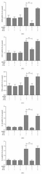
          1,8-Cineole (eucalyptol), a monoterpene, has been widely reported for the anti-inflammatory effects. Our previous data confirmed that 1,8-cineole ameliorated the inflammatory phenotype of human umbilical vein endothelial cells (HUVECs) by mediating NF-κB expression in vitro. At present, we investigated the protection effects of 1,8-cineole on vascular endothelium in lipopolysaccharide (LPS)-induced acute inflammatory injury mice and the potential mechanisms involved in the protection in HUVECs. Results from enzyme linked immunosorbent assays revealed that 1,8-cineole suppressed the secretion of interleukin (IL)-6 and IL-8 and increased the expression of IL-10 in the serum of LPS-induced mice. 1,8-Cineole reduced the inflammatory infiltration and the expression of vascular cell adhesion molecular 1 (VCAM-1) in the sections of thoracic aorta in LPS-induced acute inflammatory mice. Western blotting indicated that 1,8-cineole significantly decreased the phosphorylation of NF-κB p65 and increased the expression of PPAR-γ in the thoracic aorta tissue. 1,8-Cineole increased the expression of PPAR-γ in LPS-induced HUVECs. 1,8-Cineole and rosiglitazone reduced the protein and mRNA levels of VCAM-1, E-selectin, IL-6, and IL-8 in LPS-induced HUVECs, which could be reversed by the action of GW9662 (inhibitor of PPAR-γ). 1,8-Cineole and rosiglitazone blocked the LPS-induced IκBα degradation and NF-κB p65 nucleus translocation, which could be reversed by the pretreatment of GW9662 or silence of PPAR-γ gene. In conclusion, 1,8-cineole attenuated LPS-induced vascular endothelial cells injury via PPAR-γ dependent modulation of NF-κB.

          Related collections

          Most cited references33

          • Record: found
          • Abstract: found
          • Article: not found

          Endothelial cell metabolism in normal and diseased vasculature.

          Higher organisms rely on a closed cardiovascular circulatory system with blood vessels supplying vital nutrients and oxygen to distant tissues. Not surprisingly, vascular pathologies rank among the most life-threatening diseases. At the crux of most of these vascular pathologies are (dysfunctional) endothelial cells (ECs), the cells lining the blood vessel lumen. ECs display the remarkable capability to switch rapidly from a quiescent state to a highly migratory and proliferative state during vessel sprouting. This angiogenic switch has long been considered to be dictated by angiogenic growth factors (eg, vascular endothelial growth factor) and other signals (eg, Notch) alone, but recent findings show that it is also driven by a metabolic switch in ECs. Furthermore, these changes in metabolism may even override signals inducing vessel sprouting. Here, we review how EC metabolism differs between the normal and dysfunctional/diseased vasculature and how it relates to or affects the metabolism of other cell types contributing to the pathology. We focus on the biology of ECs in tumor blood vessel and diabetic ECs in atherosclerosis as examples of the role of endothelial metabolism in key pathological processes. Finally, current as well as unexplored EC metabolism-centric therapeutic avenues are discussed. © 2015 American Heart Association, Inc.
            Bookmark
            • Record: found
            • Abstract: found
            • Article: found
            Is Open Access

            NF-κB Signaling in Gastric Cancer

            Gastric cancer is a leading cause of cancer death worldwide. Diet, obesity, smoking and chronic infections, especially with Helicobacter pylori, contribute to stomach cancer development. H. pylori possesses a variety of virulence factors including encoded factors from the cytotoxin-associated gene pathogenicity island (cagPAI) or vacuolating cytotoxin A (VacA). Most of the cagPAI-encoded products form a type 4 secretion system (T4SS), a pilus-like macromolecular transporter, which translocates CagA into the cytoplasm of the host cell. Only H. pylori strains carrying the cagPAI induce the transcription factor NF-κB, but CagA and VacA are dispensable for direct NF-κB activation. NF-κB-driven gene products include cytokines/chemokines, growth factors, anti-apoptotic factors, angiogenesis regulators and metalloproteinases. Many of the genes transcribed by NF-κB promote gastric carcinogenesis. Since it has been shown that chemotherapy-caused cellular stress could elicit activation of the survival factor NF-κB, which leads to acquisition of chemoresistance, the NF-κB system is recommended for therapeutic targeting. Research is motivated for further search of predisposing conditions, diagnostic markers and efficient drugs to improve significantly the overall survival of patients. In this review, we provide an overview about mechanisms and consequences of NF-κB activation in gastric mucosa in order to understand the role of NF-κB in gastric carcinogenesis.
              Bookmark
              • Record: found
              • Abstract: found
              • Article: found
              Is Open Access

              BHBA Suppresses LPS-Induced Inflammation in BV-2 Cells by Inhibiting NF-κB Activation

              β -Hydroxybutyric acid (BHBA) has neuroprotective effects, but the underlying molecular mechanisms are unclear. Microglial activation plays an important role in neurodegenerative diseases by producing several proinflammatory enzymes and proinflammatory cytokines. The current study investigates the potential mechanisms whereby BHBA affects the expression of potentially proinflammatory proteins by cultured murine microglial BV-2 cells stimulated with lipopolysaccharide (LPS). The results showed that BHBA significantly reduced LPS-induced protein and mRNA expression levels of iNOS, COX-2, TNF- α , IL-1 β , and IL-6. Blocking of GPR109A by PTX resulted in a loss of this anti-inflammatory effect in BV-2 cells. Western blot analysis showed that BHBA reduced LPS-induced degradation of I κ B- α and translocation of NF- κ B, while no effect was observed on MAPKs phosphorylation. All results imply that BHBA significantly reduces levels of proinflammatory enzymes and proinflammatory cytokines by inhibition of the NF- κ B signaling pathway but not MAPKs pathways, and GPR109A is essential to this function. Overall, these data suggest that BHBA has a potential as neuroprotective drug candidate in neurodegenerative diseases.
                Bookmark

                Author and article information

                Contributors
                Journal
                Front Pharmacol
                Front Pharmacol
                Front. Pharmacol.
                Frontiers in Pharmacology
                Frontiers Media S.A.
                1663-9812
                07 March 2019
                2019
                : 10
                : 178
                Affiliations
                [1] 1The Department of Pharmacology of Materia Medica (the State Key Laboratory of Functions and Applications of Medicinal Plants, the High Efficacy Application of Natural Medicinal Resources Engineering Center of Guizhou Province, the Key Laboratory of Optimal Utilization of Natural Medicine Resources), School of Pharmaceutical Sciences, Guizhou Medical University , Guiyang, China
                [2] 2Institute of Chinese Medical Sciences, State Key Laboratory of Quality Research in Chinese Medicine, University of Macau , Macau, China
                [3] 3The Department of Pharmaceutics of TCM (the High Educational Key Laboratory of Guizhou Province for Natural Medicinal Pharmacology and Druggability, the Union Key Laboratory of Guiyang City-Guizhou Medical University), School of Pharmaceutical Sciences, Guizhou Medical University , Guiyang, China
                Author notes

                Edited by: Chiara Bolego, Università di Padova, Italy

                Reviewed by: Mingxiang Zhang, Shandong University, China; Soon Yew Tang, University of Pennsylvania, United States

                *Correspondence: Ling Tao, 649511230@ 123456qq.com Xiang-Chun Shen, shenxiangchun@ 123456126.com

                These authors have contributed equally to this work

                This article was submitted to Inflammation Pharmacology, a section of the journal Frontiers in Pharmacology

                Article
                10.3389/fphar.2019.00178
                6423908
                30930772
                6cc7c3d3-03c6-4ead-9824-069ac1470aed
                Copyright © 2019 Linghu, Wu, Fu, Yang, Li, Chen, Yu, Tao and Shen.

                This is an open-access article distributed under the terms of the Creative Commons Attribution License (CC BY). The use, distribution or reproduction in other forums is permitted, provided the original author(s) and the copyright owner(s) are credited and that the original publication in this journal is cited, in accordance with accepted academic practice. No use, distribution or reproduction is permitted which does not comply with these terms.

                History
                : 12 November 2018
                : 11 February 2019
                Page count
                Figures: 9, Tables: 1, Equations: 0, References: 40, Pages: 11, Words: 0
                Categories
                Pharmacology
                Original Research

                Pharmacology & Pharmaceutical medicine
                1,8-cineole,human umbilical vein endothelial cell,lipopolysaccharide,nf-κb,ppar-γ

                Comments

                Comment on this article