Inviting an author to review:
Find an author and click ‘Invite to review selected article’ near their name.
Search for authorsSearch for similar articles
12
views
0
recommends
+1 Recommend
0 collections
    0
    shares
      • Record: found
      • Abstract: found
      • Article: found
      Is Open Access

      Auxiliary diagnostic potential of ventricle geometry and late gadolinium enhancement in left ventricular non-compaction; non-randomized case control study

      research-article

      Read this article at

      Bookmark
          There is no author summary for this article yet. Authors can add summaries to their articles on ScienceOpen to make them more accessible to a non-specialist audience.

          Abstract

          Background

          There are still ambiguities existing in regard to left ventricular non-compaction (LVNC) diagnostic imaging. The aim of our study was to analyze diagnostic potential of late gadolinium enhancement (LGE) and ventricle geometry in patients with LVNC and controls.

          Methods

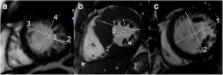
          Data on cardiac magnetic resonance imaging (CMR) studies for LVNC were reassessed from the hospital’s database (3.75 years; n=1975 exams). Matching sample of controls included cases with no structural heart disease, hypertrophic or dilative cardiomyopathy, arrhythmogenic right ventricular dysplasia or subacute myocarditis. Eccentricity of the left ventricle was measured at end diastole in the region with pronounced NC and maximal to minimal ratio (MaxMinEDDR) was calculated.

          Results

          Study included 255 patients referred for CMR, 100 (39.2%) with LVNC (prevalence in the studied period 5.01%) and 155 (60.8%) controls. Existing LGE had sensitivity of 52.5% (95%-CI:42.3–62.5), specificity of 80.4% (95%-CI:73.2–86.5) for LVNC, area under curve (AUC) 0.664 (95%-CI:0.603–0.722); p<0.001. MaxMinEDDR>1.10 had sensitivity of 95.0% (95%-CI:88.7–98.4), specificity of 82.6% (95%-CI: 75.7–88.2) for LVNC, AUC 0.917 (95%-CI:0.876–0.948); p<0.001. LGE correlated with Max-Min-EDD-R (Rho=0.130; p=0.038) and there was significant difference in ROC analysis ΔAUC0.244 (95%-CI:0.175–0.314); p<0.001. LGE also correlated negatively with stroke volume and systolic function (both p<0.05, respectively).

          Conclusions

          LGE was found to be frequently expressed in patients with LVNC, but without sufficient power to be used as a discriminative diagnostic parameter. Both LGE and eccentricity of the left ventricle were found to be relatively solid diagnostic landmarks of complex infrastructural and functional changes within the failing heart.

          Related collections

          Most cited references20

          • Record: found
          • Abstract: found
          • Article: not found

          Long-term follow-up of 34 adults with isolated left ventricular noncompaction: a distinct cardiomyopathy with poor prognosis.

          We sought to describe characteristics and outcome in adults with isolated ventricular noncompaction (IVNC). Isolated ventricular noncompaction is an unclassified cardiomyopathy due to intrauterine arrest of compaction of the loose interwoven meshwork. Knowledge regarding diagnosis, morbidity and prognosis is limited. Echocardiographic criteria for IVNC include-in the absence of significant heart lesions-segmental thickening of the left ventricular myocardial wall consisting of two layers: a thin, compacted epicardial and an extremely thickened endocardial layer with prominent trabeculations and deep recesses. Thirty-four adults (age >16 years, 25 men) fulfilled the diagnostic criteria and were followed prospectively. At diagnosis, mean age was 42 + 17 years, and 12 patients (35%) were in New York Heart Association class III/IV. Left ventricular end-diastolic diameter was 65 + 12 mm and ejection fraction 33 + 13%. Apex and/or midventricular segments of both the inferior and lateral wall were involved in >80% of patients. Follow-up was 44 + 40 months. Major complications were heart failure in 18 patients (53%), thromboembolic events in 8 patients (24%) and ventricular tachycardias in 14 patients (41%). There were 12 deaths: sudden in six, end-stage heart failure in four and other causes in two patients. Four patients underwent heart transplantation. Automated cardioverter/defibrillators were implanted in four patients. Diagnosis of IVNC by echocardiography using strict criteria is feasible. Its mortality and morbidity are high, including heart failure, thrombo-embolic events and ventricular arrhythmias. Risk stratification includes heart failure therapy, oral anticoagulation, heart transplantation and implantation of an automated defibrillator/cardioverter. As IVNC is a distinct entity, its classification as a specific cardiomyopathy seems to be more appropriate.
            Bookmark
            • Record: found
            • Abstract: found
            • Article: not found

            The pathophysiology of heart failure.

            Heart failure is a clinical syndrome that results when the heart is unable to provide sufficient blood flow to meet metabolic requirements or accommodate systemic venous return. This common condition affects over 5 million people in the United States at a cost of $10-38 billion per year. Heart failure results from injury to the myocardium from a variety of causes including ischemic heart disease, hypertension, and diabetes. Less common etiologies include cardiomyopathies, valvular disease, myocarditis, infections, systemic toxins, and cardiotoxic drugs. As the heart fails, patients develop symptoms which include dyspnea from pulmonary congestion, and peripheral edema and ascites from impaired venous return. Constitutional symptoms such as nausea, lack of appetite, and fatigue are also common. There are several compensatory mechanisms that occur as the failing heart attempts to maintain adequate function. These include increasing cardiac output via the Frank-Starling mechanism, increasing ventricular volume and wall thickness through ventricular remodeling, and maintaining tissue perfusion with augmented mean arterial pressure through activation of neurohormonal systems. Although initially beneficial in the early stages of heart failure, all of these compensatory mechanisms eventually lead to a vicious cycle of worsening heart failure. Treatment strategies have been developed based upon the understanding of these compensatory mechanisms. Medical therapy includes diuresis, suppression of the overactive neurohormonal systems, and augmentation of contractility. Surgical options include ventricular resynchronization therapy, surgical ventricular remodeling, ventricular assist device implantation, and heart transplantation. Despite significant understanding of the underlying pathophysiological mechanisms in heart failure, this disease causes significant morbidity and carries a 50% 5-year mortality. Copyright © 2012 Elsevier Inc. All rights reserved.
              Bookmark
              • Record: found
              • Abstract: found
              • Article: not found

              Measurement of trabeculated left ventricular mass using cardiac magnetic resonance imaging in the diagnosis of left ventricular non-compaction.

              To describe a method for measuring trabeculated left ventricular (LV) mass using cardiac magnetic resonance imaging and to assess its value in the diagnosis of left ventricular non-compaction (LVNC). Between January 2003 and 2008, we prospectively included 16 patients with LVNC. During the mean period, we included 16 patients with dilated cardiomyopathy (DCM), 16 patients with hypertrophic cardiomyopathy (HCM), and 16 control subjects. Left ventricular volumes, LV ejection fraction, and trabeculated LV mass were measured in the four different populations. The percentage of trabeculated LV mass was almost three times higher in the patients with LVNC (32 +/- 10%), compared with those with DCM (11 +/- 4%, P < 0.0001), HCM (12 +/- 4%, P < 0.0001), and controls (12 +/- 5%, P < 0.0001). A value of trabeculated LV mass above 20% of the global mass of the LV predicted the diagnosis of LVNC with a sensitivity of 93.7% [95% confidence interval (CI), 71.6-98.8%] and a specificity of 93.7% (95% CI, 83.1-97.8%; kappa = 0.84). The method described is reproducible and provides an assessment of the global amount of LV trabeculation. A trabeculated LV mass above 20% of the global LV mass is highly sensitive and specific for the diagnosis of LVNC.
                Bookmark

                Author and article information

                Contributors
                385 98 414 281 , marcoboban@yahoo.com
                vladimir.pesa@gmail.com
                idgabric@gmail.com
                sime.manola@kbcsm.hr
                thalassotherapiaopatia@gmail.com
                helena.antic.kauzlaric5@gmail.com
                mzulj@mefos.hr
                avcev@mefos.hr
                Journal
                BMC Cardiovasc Disord
                BMC Cardiovasc Disord
                BMC Cardiovascular Disorders
                BioMed Central (London )
                1471-2261
                6 December 2017
                6 December 2017
                2017
                : 17
                : 286
                Affiliations
                [1 ]ISNI 0000 0001 2236 1630, GRID grid.22939.33, Department of Cardiology, , University hospital “Thalassotherapia Opatija”, Faculty of Medicine, University of Rijeka, ; M. Tita 188/1, 51 440 Opatija, Croatia
                [2 ]ISNI 0000 0001 1015 399X, GRID grid.412680.9, Department of Internal medicine, , Faculty of Medicine, “J.J. Strossmayer” University of Osijek, ; Osijek, Croatia
                [3 ]ISNI 0000 0000 9336 4196, GRID grid.412488.3, Department of Cardiology, , University Hospital “Sestre Milosrdnice” Zagreb, ; Zagreb, Croatia
                Author information
                http://orcid.org/0000-0002-6129-575X
                Article
                721
                10.1186/s12872-017-0721-0
                5718034
                29207943
                6d32f8a4-2d7d-4b53-bc26-d60ad24c123a
                © The Author(s). 2017

                Open AccessThis article is distributed under the terms of the Creative Commons Attribution 4.0 International License ( http://creativecommons.org/licenses/by/4.0/), which permits unrestricted use, distribution, and reproduction in any medium, provided you give appropriate credit to the original author(s) and the source, provide a link to the Creative Commons license, and indicate if changes were made. The Creative Commons Public Domain Dedication waiver ( http://creativecommons.org/publicdomain/zero/1.0/) applies to the data made available in this article, unless otherwise stated.

                History
                : 26 August 2017
                : 27 November 2017
                Categories
                Research Article
                Custom metadata
                © The Author(s) 2017

                Cardiovascular Medicine
                left ventricle non-compaction (lvnc),late gadolinium enhancement (lge),cardiac magnetic resonance imaging (cmr),left ventricular geometry

                Comments

                Comment on this article